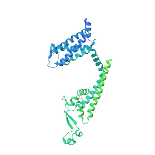
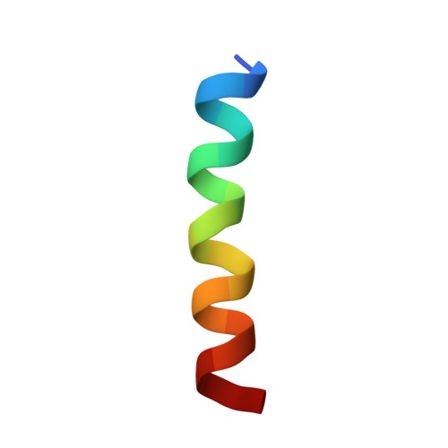
8J7H | pdb_00008j7h

ion channel


Experimental Data Snapshot

  • Method: ELECTRON MICROSCOPY
  • Resolution: 3.30 Å
  • Aggregation State: PARTICLE 
  • Reconstruction Method: SINGLE PARTICLE 

wwPDB Validation 3D Report Full Report

Validation slider image for 8J7H

This is version 1.2 of the entry. See complete history. 

Literature

Structural mechanism of voltage-gated sodium channel slow inactivation.

Chen, H., Xia, Z., Dong, J., Huang, B., Zhang, J., Zhou, F., Yan, R., Shi, Y., Gong, J., Jiang, J., Huang, Z., Jiang, D.

(2024) Nat Commun 15: 3691-3691

  • DOI: https://doi.org/10.1038/s41467-024-48125-3
  • Primary Citation Related Structures: 
    8J7F, 8J7H, 8J7M

  • PubMed Abstract: 

    Voltage-gated sodium (Na V ) channels mediate a plethora of electrical activities. Na V channels govern cellular excitability in response to depolarizing stimuli. Inactivation is an intrinsic property of Na V channels that regulates cellular excitability by controlling the channel availability. The fast inactivation, mediated by the Ile-Phe-Met (IFM) motif and the N-terminal helix (N-helix), has been well-characterized. However, the molecular mechanism underlying Na V channel slow inactivation remains elusive. Here, we demonstrate that the removal of the N-helix of Na V Eh (Na V Eh ΔN ) results in a slow-inactivated channel, and present cryo-EM structure of Na V Eh ΔN in a potential slow-inactivated state. The structure features a closed activation gate and a dilated selectivity filter (SF), indicating that the upper SF and the inner gate could serve as a gate for slow inactivation. In comparison to the Na V Eh structure, Na V Eh ΔN undergoes marked conformational shifts on the intracellular side. Together, our results provide important mechanistic insights into Na V channel slow inactivation.


  • Organizational Affiliation: 
    • Department of Microbiology and Biotechnology, College of Life Sciences, Northeast Agricultural University, No. 600 Changjiang Road, Xiangfang District, Harbin, 150030, China.

Macromolecule Content 

  • Total Structure Weight: 368.93 kDa 
  • Atom Count: 9,457 
  • Modeled Residue Count: 1,180 
  • Deposited Residue Count: 3,280 
  • Unique protein chains: 2

Macromolecules

Find similar proteins by:|  3D Structure
Entity ID: 1
MoleculeChains  Sequence LengthOrganismDetailsImage
ion channel
A, B, C, D
815Homo sapiensMutation(s): 0 
Entity Groups
Sequence Clusters30% Identity50% Identity70% Identity90% Identity95% Identity100% Identity
Sequence Annotations
Expand
Reference Sequence
Find similar proteins by:  Sequence   |   3D Structure  
Entity ID: 2
MoleculeChains  Sequence LengthOrganismDetailsImage
ILE-ALA-ALA-ILE-HIS-ASN-ALA-ARG-ARG-LYS-LYS-ARG-GLU-ALA-ALA-ALA-ALA-HIS-LYS-ALA20Homo sapiensMutation(s): 0 
Entity Groups
Sequence Clusters30% Identity50% Identity70% Identity90% Identity95% Identity100% Identity
Sequence Annotations
Expand
Reference Sequence

Experimental Data & Validation

Experimental Data

  • Method: ELECTRON MICROSCOPY
  • Resolution: 3.30 Å
  • Aggregation State: PARTICLE 
  • Reconstruction Method: SINGLE PARTICLE 
EM Software:
TaskSoftware PackageVersion
MODEL REFINEMENTPHENIX

Structure Validation

View Full Validation Report


Entry History 

& Funding Information

Deposition Data


Funding OrganizationLocationGrant Number
National Natural Science Foundation of China (NSFC)China31971134

Revision History  (Full details and data files)

  • Version 1.0: 2024-05-01
    Type: Initial release
  • Version 1.1: 2025-01-15
    Changes: Data collection, Database references, Structure summary
  • Version 1.2: 2025-07-02
    Changes: Data collection