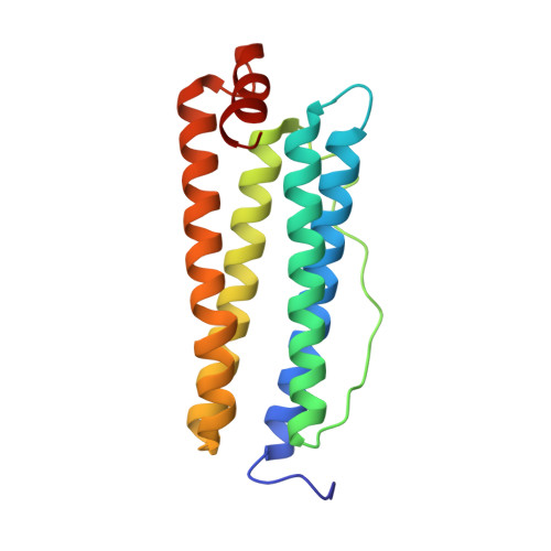
8J5A

Single-particle cryo-EM structure of mouse apoferritin at 1.19 Angstrom resolution (Dataset A)


Experimental Data Snapshot

  • Method: ELECTRON MICROSCOPY
  • Resolution: 1.19 Å
  • Aggregation State: PARTICLE 
  • Reconstruction Method: SINGLE PARTICLE 

wwPDB Validation   3D Report Full Report


This is version 1.1 of the entry. See complete history


Literature

Measurement of charges and chemical bonding in a cryo-EM structure.

Maki-Yonekura, S.Kawakami, K.Takaba, K.Hamaguchi, T.Yonekura, K.

(2023) Commun Chem 6: 98-98

  • DOI: https://doi.org/10.1038/s42004-023-00900-x
  • Primary Citation of Related Structures:  
    8J5A

  • PubMed Abstract: 

    Hydrogen bonding, bond polarity, and charges in protein molecules play critical roles in the stabilization of protein structures, as well as affecting their functions such as enzymatic catalysis, electron transfer, and ligand binding. These effects can potentially be measured in Coulomb potentials using cryogenic electron microscopy (cryo-EM). We here present charges and bond properties of hydrogen in a sub-1.2 Å resolution structure of a protein complex, apoferritin, by single-particle cryo-EM. A weighted difference map reveals positive densities for most hydrogen atoms in the core region of the complex, while negative densities around acidic amino-acid side chains are likely related to negative charges. The former positive densities identify the amino- and oxo-termini of asparagine and glutamine side chains. The latter observations were verified by spatial-resolution selection and a dose-dependent frame series. The average position of the hydrogen densities depends on the parent bonded-atom type, and this is validated by the estimated level of the standard uncertainties in the bond lengths.


  • Organizational Affiliation

    Biostructural Mechanism Laboratory, RIKEN SPring-8 Center, 1-1-1 Kouto, Sayo, Hyogo, 679-5148, Japan.


Macromolecules
Find similar proteins by:  (by identity cutoff)  |  3D Structure
Entity ID: 1
MoleculeChains Sequence LengthOrganismDetailsImage
Ferritin heavy chain172Mus musculusMutation(s): 0 
Gene Names: Fth1Fth
UniProt & NIH Common Fund Data Resources
Find proteins for P09528 (Mus musculus)
Explore P09528 
Go to UniProtKB:  P09528
IMPC:  MGI:95588
Entity Groups  
Sequence Clusters30% Identity50% Identity70% Identity90% Identity95% Identity100% Identity
UniProt GroupP09528
Sequence Annotations
Expand
  • Reference Sequence
Small Molecules
Ligands 1 Unique
IDChains Name / Formula / InChI Key2D Diagram3D Interactions
NA (Subject of Investigation/LOI)
Query on NA

Download Ideal Coordinates CCD File 
B [auth A]SODIUM ION
Na
FKNQFGJONOIPTF-UHFFFAOYSA-N
Experimental Data & Validation

Experimental Data

  • Method: ELECTRON MICROSCOPY
  • Resolution: 1.19 Å
  • Aggregation State: PARTICLE 
  • Reconstruction Method: SINGLE PARTICLE 
EM Software:
TaskSoftware PackageVersion
MODEL REFINEMENTREFMAC5.8.0267
RECONSTRUCTIONRELION3.1

Structure Validation

View Full Validation Report


Entry History & Funding Information

Deposition Data


Funding OrganizationLocationGrant Number
Japan Society for the Promotion of Science (JSPS)JapanJPMJMI20G5
Japan Society for the Promotion of Science (JSPS)JapanJPMJCR18J2

Revision History  (Full details and data files)

  • Version 1.0: 2023-07-12
    Type: Initial release
  • Version 1.1: 2024-07-03
    Changes: Data collection