8J3S | pdb_00008j3s

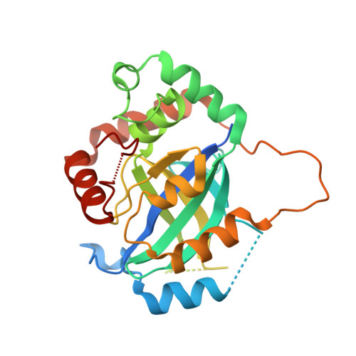
Complex structure of human cytomegalovirus protease and a macrocyclic peptide ligand


Experimental Data Snapshot

  • Method: X-RAY DIFFRACTION
  • Resolution: 3.09 Å
  • R-Value Free: 
    0.289 (Depositor), 0.284 (DCC) 
  • R-Value Work: 
    0.223 (Depositor), 0.222 (DCC) 
  • R-Value Observed: 
    0.226 (Depositor) 

wwPDB Validation 3D Report Full Report

Validation slider image for 8J3S

This is version 1.1 of the entry. See complete history

Literature

Peptide-to-Small Molecule: Discovery of Non-Covalent, Active-Site Inhibitors of beta-Herpesvirus Proteases.

Yoshida, S.Sako, Y.Nikaido, E.Ueda, T.Kozono, I.Ichihashi, Y.Nakahashi, A.Onishi, M.Yamatsu, Y.Kato, T.Nishikawa, J.Tachibana, Y.

(2023) ACS Med Chem Lett 14: 1558-1566

  • DOI: https://doi.org/10.1021/acsmedchemlett.3c00359
  • Primary Citation Related Structures: 
    8J3S, 8J3T

  • PubMed Abstract: 

    Viral proteases, the key enzymes that regulate viral replication and assembly, are promising targets for antiviral drug discovery. Herpesvirus proteases are enzymes with no crystallographically confirmed noncovalent active-site binders, owing to their shallow and polar substrate-binding pockets. Here, we applied our previously reported "Peptide-to-Small Molecule" strategy to generate novel inhibitors of β-herpesvirus proteases. Rapid selection with a display technology was used to identify macrocyclic peptide 1 bound to the active site of human cytomegalovirus protease (HCMV Pro ) with high affinity, and pharmacophore queries were defined based on the results of subsequent intermolecular interaction analyses. Membrane-permeable small molecule 19 , designed de novo according to this hypothesis, exhibited enzyme inhibitory activity (IC 50 = 10 -6 to 10 -7 M) against β-herpesvirus proteases, and the design concept was proved by X-ray cocrystal analysis.


  • Organizational Affiliation
    • Pharmaceutical Research Division, Shionogi Pharmaceutical Research Center, 3-1-1 Futaba-cho, Toyonaka, Osaka 561-0825, Japan.

Macromolecule Content 

  • Total Structure Weight: 121.62 kDa 
  • Atom Count: 6,452 
  • Modeled Residue Count: 872 
  • Deposited Residue Count: 1,091 
  • Unique protein chains: 2

Macromolecules

Find similar proteins by:|  3D Structure
Entity ID: 1
MoleculeChains  Sequence LengthOrganismDetailsImage
Assemblin
A, B, C, D
263Human betaherpesvirus 5Mutation(s): 1 
Gene Names: UL80
EC: 3.4.21.97
UniProt
Find proteins for P16753 (Human cytomegalovirus (strain AD169))
Explore P16753 
Go to UniProtKB:  P16753
Entity Groups
Sequence Clusters30% Identity50% Identity70% Identity90% Identity95% Identity100% Identity
UniProt GroupP16753
Sequence Annotations
Expand
Reference Sequence
Find similar proteins by:  Sequence   |   3D Structure  
Entity ID: 2
MoleculeChains  Sequence LengthOrganismDetailsImage
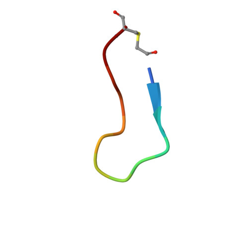
PHE-ILE-THR-GLY-HIS-TYR-TRP-VAL-ARG-PHE-LEU-PRO-CYS-GLY
E, F, G
13synthetic constructMutation(s): 0 

Small Molecules

Modified Residues  1 Unique
IDChains TypeFormula2D DiagramParent
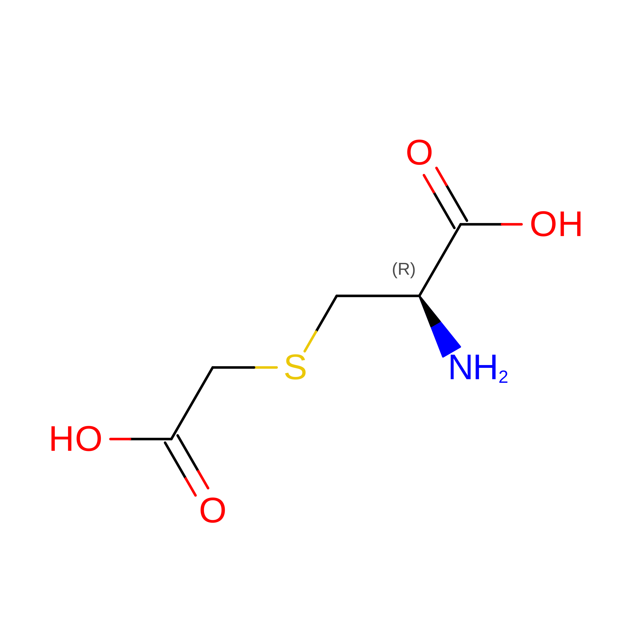
CCS
Query on CCS
E, F, G
L-PEPTIDE LINKINGC5 H9 N O4 SCYS

Experimental Data & Validation

Experimental Data

  • Method: X-RAY DIFFRACTION
  • Resolution: 3.09 Å
  • R-Value Free:  0.289 (Depositor), 0.284 (DCC) 
  • R-Value Work:  0.223 (Depositor), 0.222 (DCC) 
  • R-Value Observed: 0.226 (Depositor) 
Space Group: C 1 2 1
Unit Cell:
Length ( Å )Angle ( ˚ )
a = 79.184α = 90
b = 81.648β = 91.13
c = 198.095γ = 90
Software Package:
Software NamePurpose
REFMACrefinement
HKL-2000data scaling
HKL-2000data reduction
MOLREPphasing

Structure Validation

View Full Validation Report


Entry History 

& Funding Information

Deposition Data


Funding OrganizationLocationGrant Number
Not funded--

Revision History  (Full details and data files)

  • Version 1.0: 2023-11-08
    Type: Initial release
  • Version 1.1: 2023-11-29
    Changes: Database references