8IW5

Crystal structure of liprin-beta H2H3 dimer


Experimental Data Snapshot

  • Method: X-RAY DIFFRACTION
  • Resolution: 1.70 Å
  • R-Value Free: 0.272 
  • R-Value Work: 0.221 
  • R-Value Observed: 0.224 

wwPDB Validation   3D Report Full Report


This is version 1.0 of the entry. See complete history


Literature

KANK1 shapes focal adhesions by orchestrating protein binding, mechanical force sensing, and phase separation.

Guo, K.Zhang, J.Huang, P.Xu, Y.Pan, W.Li, K.Chen, L.Luo, L.Yu, W.Chen, S.He, S.Wei, Z.Yu, C.

(2023) Cell Rep 42: 113321-113321

  • DOI: https://doi.org/10.1016/j.celrep.2023.113321
  • Primary Citation of Related Structures:  
    8IVZ, 8IW0, 8IW5

  • PubMed Abstract: 

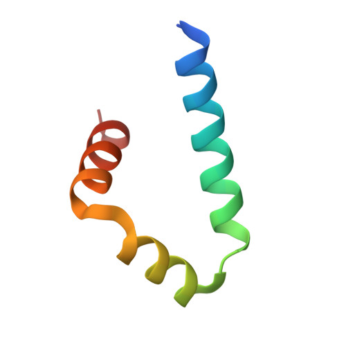
    Focal adhesions (FAs) are dynamic protein assemblies that connect cytoskeletons to the extracellular matrix and are crucial for cell adhesion and migration. KANKs are scaffold proteins that encircle FAs and act as key regulators of FA dynamics, but the molecular mechanism underlying their specified localization and functions remains poorly understood. Here, we determine the KANK1 structures in complex with talin and liprin-β, respectively. These structures, combined with our biochemical and cellular analyses, demonstrate how KANK1 scaffolds the FA core and associated proteins to modulate the FA shape in response to mechanical force. Additionally, we find that KANK1 undergoes liquid-liquid phase separation (LLPS), which is important for its localization at the FA edge and cytoskeleton connections to FAs. Our findings not only indicate the molecular basis of KANKs in bridging the core and periphery of FAs but also provide insights into the LLPS-mediated dynamic regulation of FA morphology.


  • Organizational Affiliation

    School of Life Sciences, Southern University of Science and Technology, Shenzhen, Guangdong 518055, China; Guangdong Provincial Key Laboratory of Cell Microenvironment and Disease Research, and Shenzhen Key Laboratory of Cell Microenvironment, Shenzhen 518055, Guangdong, China.


Macromolecules
Find similar proteins by:  (by identity cutoff)  |  3D Structure
Entity ID: 1
MoleculeChains Sequence LengthOrganismDetailsImage
Liprin-beta-1
A, B
51Mus musculusMutation(s): 0 
Gene Names: Ppfibp1Kiaa1230
UniProt
Find proteins for Q8C8U0 (Mus musculus)
Explore Q8C8U0 
Go to UniProtKB:  Q8C8U0
Entity Groups  
Sequence Clusters30% Identity50% Identity70% Identity90% Identity95% Identity100% Identity
UniProt GroupQ8C8U0
Sequence Annotations
Expand
  • Reference Sequence
Small Molecules
Ligands 1 Unique
IDChains Name / Formula / InChI Key2D Diagram3D Interactions
CA
Query on CA

Download Ideal Coordinates CCD File 
C [auth B]CALCIUM ION
Ca
BHPQYMZQTOCNFJ-UHFFFAOYSA-N
Experimental Data & Validation

Experimental Data

  • Method: X-RAY DIFFRACTION
  • Resolution: 1.70 Å
  • R-Value Free: 0.272 
  • R-Value Work: 0.221 
  • R-Value Observed: 0.224 
  • Space Group: P 1 21 1
Unit Cell:
Length ( Å )Angle ( ˚ )
a = 29.793α = 90
b = 50.637β = 113.44
c = 30.78γ = 90
Software Package:
Software NamePurpose
PHENIXrefinement
SCALEPACKdata scaling
PDB_EXTRACTdata extraction
HKL-2000data reduction
PHENIXphasing

Structure Validation

View Full Validation Report


Entry History & Funding Information

Deposition Data


Funding OrganizationLocationGrant Number
National Natural Science Foundation of China (NSFC)China31971131
National Natural Science Foundation of China (NSFC)China32170697

Revision History  (Full details and data files)

  • Version 1.0: 2023-11-08
    Type: Initial release