Klebsiella pneumoniae K2 capsular polysaccharide degradation by a bacteriophage depolymerase does not require trimer formation.
Ye, T.J., Fung, K.M., Lee, I.M., Ko, T.P., Lin, C.Y., Wong, C.L., Tu, I.F., Huang, T.Y., Yang, F.L., Chang, Y.P., Wang, J.T., Lin, T.L., Huang, K.F., Wu, S.H.(2024) mBio 15: e0351923-e0351923
- PubMed: 38349137 Search on PubMedSearch on PubMed Central
- DOI: https://doi.org/10.1128/mbio.03519-23
- Primary Citation Related Structures:
8IQ5, 8IQ9, 8IQE - PubMed Abstract:
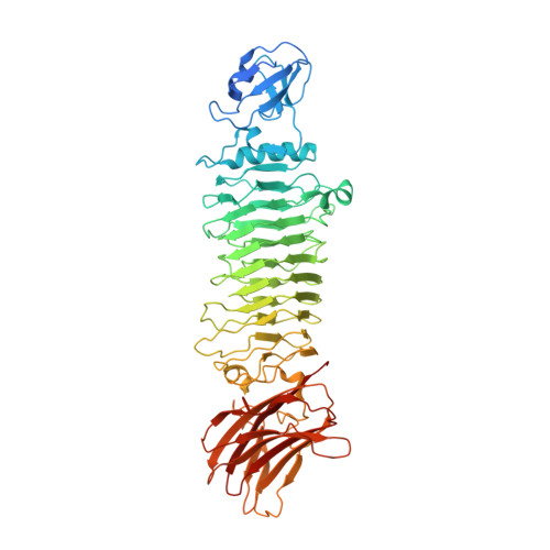
K2-capsular Klebsiella pneumoniae is a hypervirulent pathogen that causes fatal infections. Here, we describe a phage tailspike protein, named K2-2, that specifically depolymerizes the K2 capsular polysaccharide (CPS) of K. pneumoniae into tetrasaccharide repeating units. Nearly half of the products contained O -acetylation, which was thought crucial to the immunogenicity of CPS. The product-bound structures of this trimeric enzyme revealed intersubunit carbohydrate-binding grooves, each accommodating three tetrasaccharide units of K2 CPS. The catalytic residues and the key interactions responsible for K2 CPS recognition were identified and verified by site-directed mutagenesis. Further biophysical and functional characterization, along with the structure of a tetrameric form of K2-2, demonstrated that the formation of intersubunit catalytic center does not require trimerization, which could be nearly completely disrupted by a single-residue mutation in the C-terminal domain. Our findings regarding the assembly and catalysis of K2-2 provide cues for the development of glycoconjugate vaccines against K. pneumoniae infection. Generating fragments of capsular polysaccharides from pathogenic bacteria with crucial antigenic determinants for vaccine development continues to pose challenges. The significance of the C-terminal region of phage tailspike protein (TSP) in relation to its folding and trimer formation remains largely unexplored. The polysaccharide depolymerase described here demonstrates the ability to depolymerize the K2 CPS of K. pneumoniae into tetrasaccharide fragments while retaining the vital O -acetylation modification crucial for immunogenicity. By carefully characterizing the enzyme, elucidating its three-dimensional structures, conducting site-directed mutagenesis, and assessing the antimicrobial efficacy of the mutant enzymes against K2 K. pneumoniae , we offer valuable insights into the mechanism by which this enzyme recognizes and depolymerizes the K2 CPS. Our findings, particularly the discovery that trimer formation is not required for depolymerizing activity, challenge the current understanding of trimer-dependent TSP activity and highlight the catalytic mechanism of the TSP with an intersubunit catalytic center.
- Institute of Biological Chemistry, Academia Sinica, Taipei, Taiwan.
Organizational Affiliation: