8IPP | pdb_00008ipp

Crystal structure of the complex between an ankyrin and a parallel G-quadruplex


Experimental Data Snapshot

  • Method: X-RAY DIFFRACTION
  • Resolution: 2.01 Å
  • R-Value Free: 
    0.224 (Depositor), 0.274 (DCC) 
  • R-Value Work: 
    0.211 (Depositor), 0.254 (DCC) 

Starting Models: experimental
View more details

wwPDB Validation 3D Report Full Report

Validation slider image for 8IPP

This is version 1.0 of the entry. See complete history

Literature

Structural Basis for Parallel G-Quadruplex Recognition by an Ankyrin Protein.

Ngo, K.H.Liew, C.W.Heddi, B.Phan, A.T.

(2024) J Am Chem Soc 146: 13709-13713

  • DOI: https://doi.org/10.1021/jacs.4c01971
  • Primary Citation Related Structures: 
    8IPF, 8IPP

  • PubMed Abstract: 

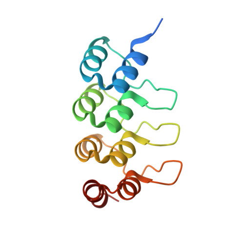
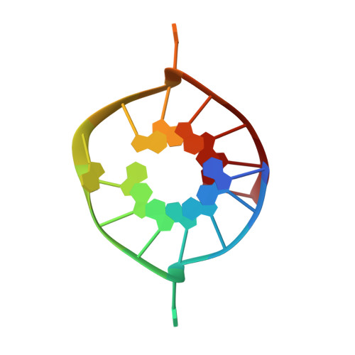
    G-Quadruplex (G4) structures formed by guanine-rich DNA and RNA sequences are implicated in various biological processes. Understanding the mechanisms by which proteins recognize G4 structures is crucial for elucidating their functional roles. Here we present the X-ray crystal structure of an ankyrin protein bound to a parallel G4 structure. Our findings reveal a new specific recognition mode in which a bundle of α-helices and loops of the ankyrin form a flat surface to stack on the G-tetrad core. The protein employs a combination of hydrogen bonds and hydrophobic contacts to interact with the G4, and electrostatic interaction is used to enhance the binding affinity. This binding mechanism provides valuable insights into understanding G4 recognition by proteins.


  • Organizational Affiliation
    • School of Physical and Mathematical Sciences, Nanyang Technological University, Singapore 637371, Singapore.

Macromolecule Content 

  • Total Structure Weight: 20.16 kDa 
  • Atom Count: 1,406 
  • Modeled Residue Count: 142 
  • Deposited Residue Count: 152 
  • Unique protein chains: 1
  • Unique nucleic acid chains: 1

Macromolecules


Find similar proteins by:|  3D Structure
Entity ID: 2
MoleculeChains  Sequence LengthOrganismDetailsImage
DARPin protein 2E4136synthetic constructMutation(s): 0 
Find similar nucleic acids by:  Sequence
Entity ID: 1
MoleculeChains LengthOrganismImage
93del-T1216synthetic construct
Sequence Annotations
Expand
Reference Sequence

Experimental Data & Validation

Experimental Data

  • Method: X-RAY DIFFRACTION
  • Resolution: 2.01 Å
  • R-Value Free:  0.224 (Depositor), 0.274 (DCC) 
  • R-Value Work:  0.211 (Depositor), 0.254 (DCC) 
Space Group: P 21 21 2
Unit Cell:
Length ( Å )Angle ( ˚ )
a = 76.759α = 90
b = 83.587β = 90
c = 31.858γ = 90
Software Package:
Software NamePurpose
REFMACrefinement
XDSdata reduction
XDSdata scaling
PHASERphasing

Structure Validation

View Full Validation Report


Entry History 

& Funding Information

Deposition Data


Funding OrganizationLocationGrant Number
National Research Foundation (NRF, Singapore)SingaporeNRF-NRFI2017-09
Ministry of Education (MoE, Singapore)SingaporeMOE2018-T2-2-029

Revision History  (Full details and data files)

  • Version 1.0: 2024-07-31
    Type: Initial release