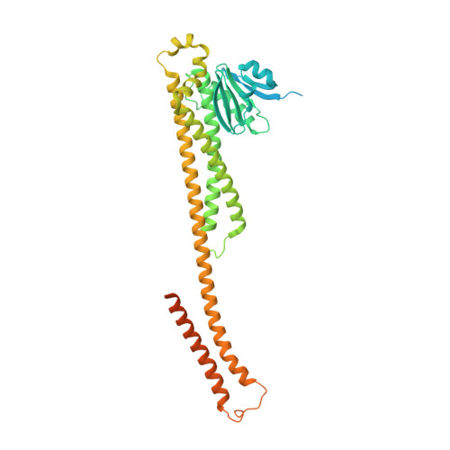
8IP4 | pdb_00008ip4

Cryo-EM structure of hMRS-highEDTA


Experimental Data Snapshot

  • Method: ELECTRON MICROSCOPY
  • Resolution: 2.70 Å
  • Aggregation State: PARTICLE 
  • Reconstruction Method: SINGLE PARTICLE 

wwPDB Validation 3D Report Full Report

Validation slider image for 8IP4

This is version 1.2 of the entry. See complete history. 

Literature

Molecular basis of Mg 2+ permeation through the human mitochondrial Mrs2 channel.

Li, M., Li, Y., Lu, Y., Li, J., Lu, X., Ren, Y., Wen, T., Wang, Y., Chang, S., Zhang, X., Yang, X., Shen, Y.

(2023) Nat Commun 14: 4713-4713

  • DOI: https://doi.org/10.1038/s41467-023-40516-2
  • Primary Citation Related Structures: 
    8IP3, 8IP4, 8IP5, 8IP6

  • PubMed Abstract: 

    Mitochondrial RNA splicing 2 (Mrs2), a eukaryotic CorA ortholog, enables Mg 2+ to permeate the inner mitochondrial membrane and plays an important role in mitochondrial metabolic function. However, the mechanism by which Mrs2 permeates Mg 2+ remains unclear. Here, we report four cryo-electron microscopy (cryo-EM) reconstructions of Homo sapiens Mrs2 (hMrs2) under various conditions. All of these hMrs2 structures form symmetrical pentamers with very similar pentamer and protomer conformations. A special structural feature of Cl - -bound R-ring, which consists of five Arg332 residues, was found in the hMrs2 structure. Molecular dynamics simulations and mitochondrial Mg 2+ uptake assays show that the R-ring may function as a charge repulsion barrier, and Cl - may function as a ferry to jointly gate Mg 2+ permeation in hMrs2. In addition, the membrane potential is likely to be the driving force for Mg 2+ permeation. Our results provide insights into the channel assembly and Mg 2+ permeation of hMrs2.


  • Organizational Affiliation: 
    • State Key Laboratory of Medicinal Chemical Biology and Frontiers Science Center for Cell Responses, College of Life Sciences, Nankai University, Tianjin, 300350, China.

Macromolecule Content 

  • Total Structure Weight: 257.74 kDa 
  • Atom Count: 12,933 
  • Modeled Residue Count: 1,600 
  • Deposited Residue Count: 2,265 
  • Unique protein chains: 1

Macromolecules

Find similar proteins by:|  3D Structure
Entity ID: 1
MoleculeChains  Sequence LengthOrganismDetailsImage
Magnesium transporter MRS2 homolog, mitochondrial
A, B, C, D, E
453Homo sapiensMutation(s): 0 
Gene Names: MRS2, HPT, MRS2L
Membrane Entity: Yes 
UniProt & NIH Common Fund Data Resources
Find proteins for Q9HD23 (Homo sapiens)
Explore Q9HD23 
Go to UniProtKB:  Q9HD23
PHAROS:  Q9HD23
GTEx:  ENSG00000124532 
Entity Groups
Sequence Clusters30% Identity50% Identity70% Identity90% Identity95% Identity100% Identity
UniProt GroupQ9HD23
Sequence Annotations
Expand
Reference Sequence

Experimental Data & Validation

Experimental Data

  • Method: ELECTRON MICROSCOPY
  • Resolution: 2.70 Å
  • Aggregation State: PARTICLE 
  • Reconstruction Method: SINGLE PARTICLE 
EM Software:
TaskSoftware PackageVersion
MODEL REFINEMENTPHENIX

Structure Validation

View Full Validation Report


Entry History 

& Funding Information

Deposition Data


Funding OrganizationLocationGrant Number
National Science Foundation (NSF, China)China--

Revision History  (Full details and data files)

  • Version 1.0: 2023-06-14
    Type: Initial release
  • Version 1.1: 2023-08-30
    Changes: Data collection, Database references
  • Version 1.2: 2025-07-23
    Changes: Data collection, Structure summary