8IN1

beta-glucosidase protein from Aplysia kurodai

  • Classification: HYDROLASE
  • Organism(s): Aplysia kurodai
  • Mutation(s): No 

  • Deposited: 2023-03-08 Released: 2023-11-15 
  • Deposition Author(s): Sun, X.M., Ye, Y.X., Kato, K., Yu, J., Yao, M.
  • Funding Organization(s): Japan Society for the Promotion of Science (JSPS), Japan Agency for Medical Research and Development (AMED)

Experimental Data Snapshot

  • Method: X-RAY DIFFRACTION
  • Resolution: 2.70 Å
  • R-Value Free: 0.220 
  • R-Value Work: 0.183 
  • R-Value Observed: 0.185 

wwPDB Validation   3D Report Full Report


Ligand Structure Quality Assessment 


This is version 1.0 of the entry. See complete history


Literature

Structural basis of EHEP-mediated offense against phlorotannin-induced defense from brown algae to protect aku BGL activity.

Sun, X.Ye, Y.Sakurai, N.Wang, H.Kato, K.Yu, J.Yuasa, K.Tsuji, A.Yao, M.

(2023) Elife 12

  • DOI: https://doi.org/10.7554/eLife.88939
  • Primary Citation of Related Structures:  
    8IN1, 8IN3, 8IN4, 8IN6

  • PubMed Abstract: 

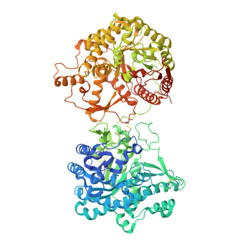
    The defensive-offensive associations between algae and herbivores determine marine ecology. Brown algae utilize phlorotannin as their chemical defense against the predator Aplysia kurodai , which uses β-glucosidase ( aku BGL) to digest the laminarin in algae into glucose. Moreover, A. kurodai employs Eisenia hydrolysis-enhancing protein (EHEP) as an offense to protect aku BGL activity from phlorotannin inhibition by precipitating phlorotannin. To underpin the molecular mechanism of this digestive-defensive-offensive system, we determined the structures of the apo and tannic acid (TNA, a phlorotannin analog) bound forms of EHEP, as well as the apo aku BGL. EHEP consisted of three peritrophin-A domains arranged in a triangular shape and bound TNA in the center without significant conformational changes. Structural comparison between EHEP and EHEP-TNA led us to find that EHEP can be resolubilized from phlorotannin precipitation at an alkaline pH, which reflects a requirement in the digestive tract. aku BGL contained two GH1 domains, only one of which conserved the active site. Combining docking analysis, we propose the mechanisms by which phlorotannin inhibits aku BGL by occupying the substrate-binding pocket, and EHEP protects aku BGL against this inhibition by binding with phlorotannin to free the aku BGL pocket.


  • Organizational Affiliation

    Faculty of Advanced Life Science, Hokkaido University, Sapporo, Japan.


Macromolecules
Find similar proteins by:  (by identity cutoff)  |  3D Structure
Entity ID: 1
MoleculeChains Sequence LengthOrganismDetailsImage
Beta-Glucosidase
A, B
994Aplysia kurodaiMutation(s): 0 
UniProt
Find proteins for A0A1V1FXL2 (Aplysia kurodai)
Explore A0A1V1FXL2 
Go to UniProtKB:  A0A1V1FXL2
Entity Groups  
Sequence Clusters30% Identity50% Identity70% Identity90% Identity95% Identity100% Identity
UniProt GroupA0A1V1FXL2
Sequence Annotations
Expand
  • Reference Sequence
Oligosaccharides

Help

Entity ID: 2
MoleculeChains Length2D Diagram Glycosylation3D Interactions
beta-D-mannopyranose-(1-4)-2-acetamido-2-deoxy-beta-D-glucopyranose-(1-4)-[alpha-L-fucopyranose-(1-3)][alpha-L-fucopyranose-(1-6)]2-acetamido-2-deoxy-beta-D-glucopyranoseC [auth D],
F [auth G]
5N-Glycosylation
Glycosylation Resources
GlyTouCan:  G92953ND
GlyCosmos:  G92953ND
Entity ID: 3
MoleculeChains Length2D Diagram Glycosylation3D Interactions
alpha-L-fucopyranose-(1-3)-2-acetamido-2-deoxy-beta-D-glucopyranoseD [auth E]2N-Glycosylation
Glycosylation Resources
GlyTouCan:  G80587NA
GlyCosmos:  G80587NA
GlyGen:  G80587NA
Entity ID: 4
MoleculeChains Length2D Diagram Glycosylation3D Interactions
beta-D-mannopyranose-(1-4)-2-acetamido-2-deoxy-beta-D-glucopyranose-(1-4)-[alpha-L-fucopyranose-(1-3)]2-acetamido-2-deoxy-beta-D-glucopyranoseE [auth F]4N-Glycosylation
Glycosylation Resources
GlyTouCan:  G18638YB
GlyCosmos:  G18638YB
GlyGen:  G18638YB
Entity ID: 5
MoleculeChains Length2D Diagram Glycosylation3D Interactions
alpha-L-fucopyranose-(1-3)-[2-acetamido-2-deoxy-beta-D-glucopyranose-(1-4)]2-acetamido-2-deoxy-beta-D-glucopyranoseG [auth I]3N-Glycosylation
Glycosylation Resources
GlyTouCan:  G11971MR
GlyCosmos:  G11971MR
GlyGen:  G11971MR
Small Molecules
Ligands 1 Unique
IDChains Name / Formula / InChI Key2D Diagram3D Interactions
NAG
Query on NAG

Download Ideal Coordinates CCD File 
H [auth B]2-acetamido-2-deoxy-beta-D-glucopyranose
C8 H15 N O6
OVRNDRQMDRJTHS-FMDGEEDCSA-N
Experimental Data & Validation

Experimental Data

  • Method: X-RAY DIFFRACTION
  • Resolution: 2.70 Å
  • R-Value Free: 0.220 
  • R-Value Work: 0.183 
  • R-Value Observed: 0.185 
  • Space Group: P 62
Unit Cell:
Length ( Å )Angle ( ˚ )
a = 191.669α = 90
b = 191.669β = 90
c = 112.551γ = 120
Software Package:
Software NamePurpose
PHENIXrefinement
XDSdata reduction
XDSdata scaling
PHENIXphasing

Structure Validation

View Full Validation Report


Ligand Structure Quality Assessment 


Entry History & Funding Information

Deposition Data


Funding OrganizationLocationGrant Number
Japan Society for the Promotion of Science (JSPS)Japan21H01754
Japan Agency for Medical Research and Development (AMED)JapanJP18am0101071
Japan Agency for Medical Research and Development (AMED)JapanJP19am0101083

Revision History  (Full details and data files)

  • Version 1.0: 2023-11-15
    Type: Initial release