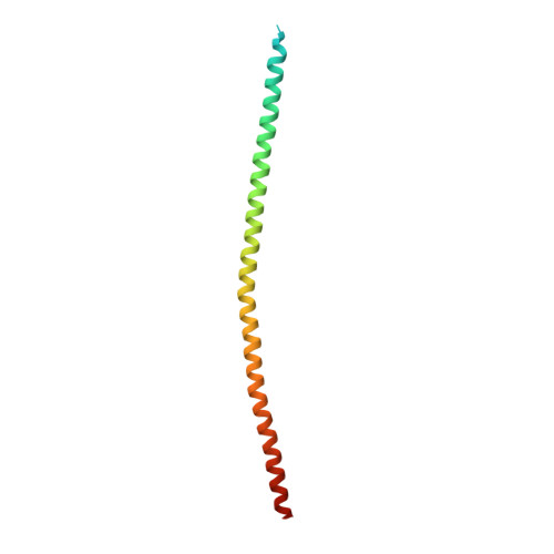
The structure of TRAF7 coiled-coil trimer provides insight into its function in zebrafish embryonic development.
Song, X., Hu, R., Chen, Y., Xiao, M., Zhang, H., Wu, S., Lu, Q.(2024) J Mol Cell Biol 16
- PubMed: 38178633 Search on PubMed
- DOI: https://doi.org/10.1093/jmcb/mjad083
- Primary Citation Related Structures:
8IMS - PubMed Abstract:
TRAF7 serves as a crucial intracellular adaptor and E3 ubiquitin ligase involved in signal transduction pathways, contributing to immune responses, tumor progression, and embryonic development. Somatic mutations within the coiled-coil (CC) domain and WD40 repeat domain of TRAF7 could cause brain tumors, while germline pathogenic mutations contribute to severe developmental abnormalities. However, the precise molecular mechanism underlying TRAF7 involvement in embryonic development remains unclear. In this study, we employed zebrafish as an in-vivo model system. TRAF7 knockdown caused defects in zebrafish embryonic development. We determined the crystal structure of TRAF7 CC domain at 3.3 Å resolution and found that the CC region trimerization was essential for TRAF7 functionality during zebrafish embryonic development. Additionally, disease-causing mutations in TRAF7 CC region could impair the trimer formation, consequently impacting early embryonic development of zebrafish. Therefore, our study sheds light on the molecular mechanism of TRAF7 CC trimer formation and its pivotal role in embryonic development.
- Molecular Diagnostic Laboratory, Shanghai Children's Hospital, School of Medicine, Shanghai Jiao Tong University, Shanghai 200040, China.
Organizational Affiliation: