8IMD | pdb_00008imd

Crystal structure of Cu/Zn Superoxide dismutase from Paenibacillus lautus


Experimental Data Snapshot

  • Method: X-RAY DIFFRACTION
  • Resolution: 1.45 Å
  • R-Value Free: 
    0.221 (Depositor), 0.221 (DCC) 
  • R-Value Work: 
    0.188 (Depositor), 0.188 (DCC) 
  • R-Value Observed: 
    0.190 (Depositor) 

wwPDB Validation 3D Report Full Report

Validation slider image for 8IMD

This is version 1.0 of the entry. See complete history

Literature

Characterization of a novel cysteine-less Cu/Zn-superoxide dismutase in Paenibacillus lautus missing a conserved disulfide bond.

Furukawa, Y.Shintani, A.Narikiyo, S.Sue, K.Akutsu, M.Muraki, N.

(2023) J Biological Chem 299: 105040-105040

  • DOI: https://doi.org/10.1016/j.jbc.2023.105040
  • Primary Citation Related Structures: 
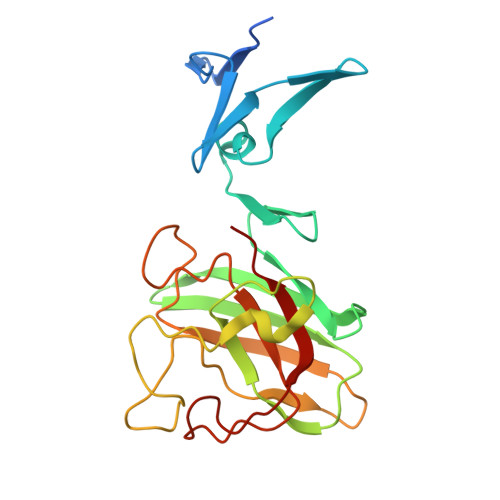
    8IMD

  • PubMed Abstract: 

    Cu/Zn-superoxide dismutase (CuZnSOD) is an enzyme that binds a copper and zinc ion and also forms an intramolecular disulfide bond. Together with the copper ion as the active site, the disulfide bond is completely conserved among these proteins; indeed, the disulfide bond plays critical roles in maintaining the catalytically competent conformation of CuZnSOD. Here, we found that a CuZnSOD protein in Paenibacillus lautus (PaSOD) has no Cys residue but exhibits a significant level of enzyme activity. The crystal structure of PaSOD revealed hydrophobic and hydrogen-bonding interactions in substitution for the disulfide bond of the other CuZnSOD proteins. Also notably, we determined that PaSOD forms a homodimer through an additional domain with a novel fold at the N terminus. While the advantages of lacking Cys residues and adopting a novel dimer configuration remain obscure, PaSOD does not require a disulfide-introducing/correcting system for maturation and could also avoid misfolding caused by aberrant thiol oxidations under an oxidative environment.


  • Organizational Affiliation
    • Department of Chemistry, Keio University, Yokohama, Japan. Electronic address: furukawa@chem.keio.ac.jp.

Macromolecule Content 

  • Total Structure Weight: 47.18 kDa 
  • Atom Count: 3,478 
  • Modeled Residue Count: 412 
  • Deposited Residue Count: 444 
  • Unique protein chains: 1

Macromolecules

Find similar proteins by:|  3D Structure
Entity ID: 1
MoleculeChains  Sequence LengthOrganismDetailsImage
Cu/Zn-Superoxide dismutase
A, B
222Paenibacillus lautus NBRC 15380Mutation(s): 0 
UniProt
Find proteins for A0AAJ6N6D8 (Paenibacillus lautus NBRC 15380)
Explore A0AAJ6N6D8 
Go to UniProtKB:  A0AAJ6N6D8
Entity Groups
Sequence Clusters30% Identity50% Identity70% Identity90% Identity95% Identity100% Identity
UniProt GroupA0AAJ6N6D8
Sequence Annotations
Expand
Reference Sequence

Small Molecules

Experimental Data & Validation

Experimental Data

  • Method: X-RAY DIFFRACTION
  • Resolution: 1.45 Å
  • R-Value Free:  0.221 (Depositor), 0.221 (DCC) 
  • R-Value Work:  0.188 (Depositor), 0.188 (DCC) 
  • R-Value Observed: 0.190 (Depositor) 
Space Group: P 21 21 21
Unit Cell:
Length ( Å )Angle ( ˚ )
a = 45.24α = 90
b = 53.612β = 90
c = 169.567γ = 90
Software Package:
Software NamePurpose
REFMACrefinement
XDSdata reduction
SCALAdata scaling
CRANK2phasing

Structure Validation

View Full Validation Report


Entry History 

& Funding Information

Deposition Data


Funding OrganizationLocationGrant Number
Ministry of Education, Culture, Sports, Science and Technology (Japan)Japan19H05765

Revision History  (Full details and data files)

  • Version 1.0: 2024-01-17
    Type: Initial release