Characterization of the molecular interactions between resveratrol derivatives and death-associated protein kinase 1.
Yokoyama, T., Kusaka, K.(2023) FEBS J 290: 4465-4479
- PubMed: 37171222 Search on PubMed
- DOI: https://doi.org/10.1111/febs.16817
- Primary Citation Related Structures:
8IE5, 8IE6, 8IE7, 8IE8 - PubMed Abstract:
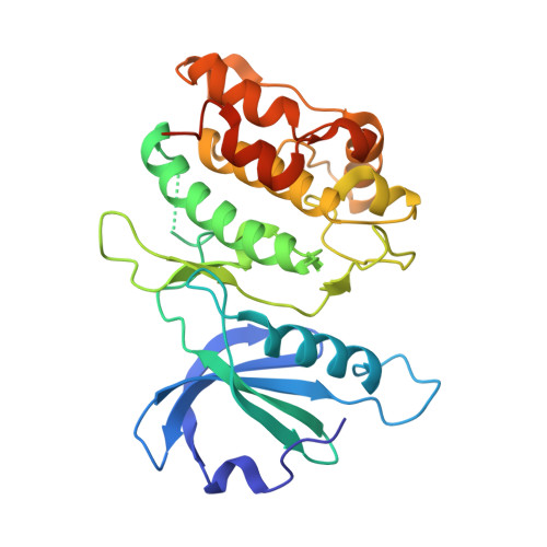
Death-associated protein kinase 1 (DAPK1), a Ca2+/calmodulin-regulated serine/threonine kinase, regulates cell apoptosis and autophagy and has been implicated in the pathogenesis of Alzheimer's disease (AD). Targeting DAPK1 may be a promising approach for treating AD. In our previous study, we found that a natural polyphenol, resveratrol (1), is a moderate DAPK1 inhibitor. In the present study, we investigated the interactions between natural and synthetic derivatives of 1 and DAPK1. Binding assays including intrinsic fluorescence quenching, protein thermal shift and isothermal titration calorimetry indicated that oxyresveratrol (3), a hydroxylated derivative, and pinostilbene (5), a methoxylated derivative, bind to DAPK1 with comparable affinity to 1. The enzymatic assay showed that 3 more effectively inhibits the intrinsic ATPase activity of DAPK1 compared with 1. Crystallographic analysis revealed that the binding modes of the methoxylated derivatives were different from those of 1 and 3, resulting in a unique interaction. Our results suggest that 3 may be helpful in treating AD and provide a clue for the development of promising DAPK1 inhibitors.
- Faculty of Pharmaceutical Sciences, University of Toyama, Japan.
Organizational Affiliation: