Recombinant human scFv antibody fragments against phospholipase A2 from Naja naja and Echis carinatus snake venoms: In vivo neutralization and mechanistic insights.
Kumar, A., Madni, Z.K., Chaturvedi, S., Salunke, D.M.(2024) Mol Immunol 165: 55-67
- PubMed: 38154407 Search on PubMed
- DOI: https://doi.org/10.1016/j.molimm.2023.12.006
- Primary Citation Related Structures:
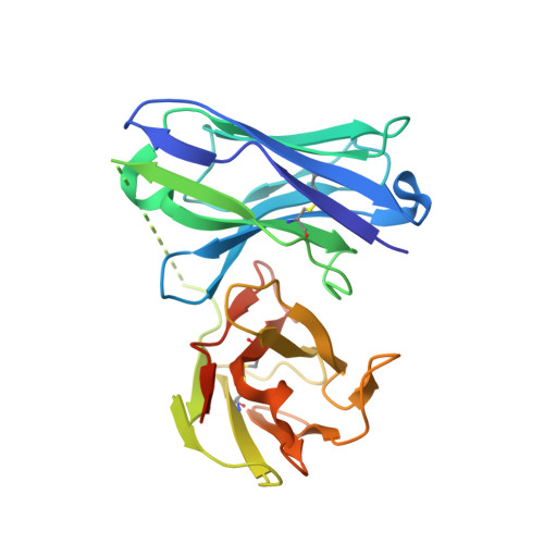
8IA6 - PubMed Abstract:
Snake envenomation results in a range of clinical sequelae, and widely used animal-based conventional antivenoms exhibit several limitations including the adverse immunological effects in human snake bite victims. Therefore, human monoclonal anti-snake venom antibodies or fragments can be an alternate therapy for overcoming the existing limitations. We developed venom-neutralizing humanized scFv antibodies and analyzed biochemical mechanisms associated with the inhibition of toxicity. Tomlinson I and J human scFv antibody libraries were screened against Naja naja and Echis carinatus venoms, and seven unique scFv antibodies were obtained. Further, specific toxins of snake venom interacting with each of these scFvs were identified, and phospholipase A2 (PLA2) was found to be prominently captured by the phage-anchored scFv antibodies. Our study indicated PLA2 to be one of the abundant toxins in Naja naja and Echis carinatus venom samples. The scFvs binding to PLA2 were used to perform in vivo survival assay using the mouse model and in vitro toxin inhibition assays. scFv N194, which binds to acidic PLA2, protected 50% of mice treated with Naja naja venom. Significant prolongation of survival time and 16% survival were observed in Echis carinatus venom-challenged mice treated with scFv E113 and scFv E10, respectively. However, a combination comprised of an equal amount of two scFvs, E113 and E10, both interacting with basic PLA2, exhibited synergistically enhanced survival of 33% in Echis carinatus venom-challenged mice. No such synergistically enhanced survival was observed in the case of combinatorial treatment with anti-Naja naja scFvs, N194, and N248. These scFvs demonstrated partial inhibition of venom-induced myotoxicity, and E113 also inhibited hemolysis by 50%, which corroborates the enhanced survival during combinatorial treatment in Echis carinatus venom-challenged mice.
- International Centre for Genetic Engineering and Biotechnology, Aruna Asaf Ali Marg, New Delhi 110067, India.
Organizational Affiliation: