8I76 | pdb_00008i76

Crystal structure of decarboxylated osteocalcin at pH 2.0 without glycerol


Experimental Data Snapshot

  • Method: X-RAY DIFFRACTION
  • Resolution: 1.38 Å
  • R-Value Free: 
    0.221 (Depositor), 0.223 (DCC) 
  • R-Value Work: 
    0.195 (Depositor), 0.196 (DCC) 
  • R-Value Observed: 
    0.196 (Depositor) 

Starting Model: experimental
View more details

wwPDB Validation 3D Report Full Report

Validation slider image for 8I76

This is version 1.2 of the entry. See complete history

Literature

Structural and mutational analyses of decarboxylated osteocalcin provide insight into its adiponectin-inducing activity.

Mizuguchi, M.Yokoyama, T.Otani, T.Kuribara, S.Nabeshima, Y.Obita, T.Hirata, M.Kawano, K.

(2023) FEBS Lett 597: 1479-1488

  • DOI: https://doi.org/10.1002/1873-3468.14618
  • Primary Citation Related Structures: 
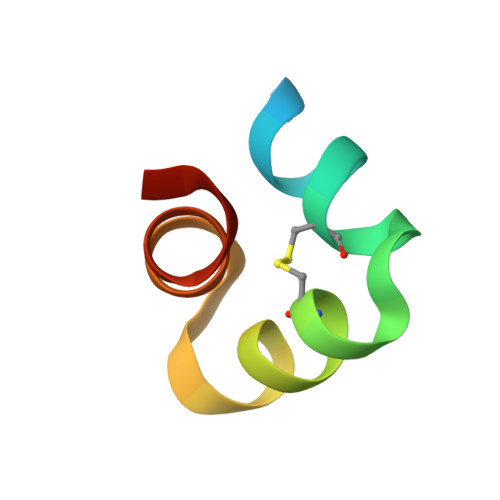
    8I74, 8I75, 8I76

  • PubMed Abstract: 

    An acidic environment in bone is essential for bone metabolism and the production of decarboxylated osteocalcin, which functions as a regulatory hormone of glucose metabolism. Here, we describe the high-resolution X-ray crystal structure of decarboxylated osteocalcin under acidic conditions. Decarboxylated osteocalcin at pH 2.0 retains the α-helix structure of native osteocalcin with three γ-carboxyglutamic acid residues at neutral pH. This implies that decarboxylated osteocalcin is stable under an acidic environment in bone. In addition, site-directed mutagenesis revealed that Glu17 and Glu21 are important for the adiponectin-inducing activity of decarboxylated osteocalcin. These findings suggest that the receptor of decarboxylated osteocalcin responds to the negative charge in helix 1 of osteocalcin.


  • Organizational Affiliation
    • Faculty of Pharmaceutical Sciences, University of Toyama, Toyama, Japan.

Macromolecule Content 

  • Total Structure Weight: 8.5 kDa 
  • Atom Count: 590 
  • Modeled Residue Count: 62 
  • Deposited Residue Count: 76 
  • Unique protein chains: 1

Macromolecules

Find similar proteins by:|  3D Structure
Entity ID: 1
MoleculeChains  Sequence LengthOrganismDetailsImage
Osteocalcin
A, B
38Bos taurusMutation(s): 0 
Gene Names: BGLAP
UniProt
Find proteins for P02820 (Bos taurus)
Explore P02820 
Go to UniProtKB:  P02820
Entity Groups
Sequence Clusters30% Identity50% Identity70% Identity90% Identity95% Identity100% Identity
UniProt GroupP02820
Sequence Annotations
Expand
Reference Sequence

Experimental Data & Validation

Experimental Data

  • Method: X-RAY DIFFRACTION
  • Resolution: 1.38 Å
  • R-Value Free:  0.221 (Depositor), 0.223 (DCC) 
  • R-Value Work:  0.195 (Depositor), 0.196 (DCC) 
  • R-Value Observed: 0.196 (Depositor) 
Space Group: P 41
Unit Cell:
Length ( Å )Angle ( ˚ )
a = 42.105α = 90
b = 42.105β = 90
c = 36.711γ = 90
Software Package:
Software NamePurpose
PHENIXrefinement
XDSdata reduction
XSCALEdata scaling
PHENIXphasing

Structure Validation

View Full Validation Report


Entry History 

& Funding Information

Deposition Data


Funding OrganizationLocationGrant Number
Not funded--

Revision History  (Full details and data files)

  • Version 1.0: 2024-01-31
    Type: Initial release
  • Version 1.1: 2024-08-21
    Changes: Database references
  • Version 1.2: 2024-11-13
    Changes: Structure summary