8I5W

Crystal structure of the DHR-2 domain of DOCK10 in complex with Rac1


Experimental Data Snapshot

  • Method: X-RAY DIFFRACTION
  • Resolution: 2.43 Å
  • R-Value Free: 0.280 
  • R-Value Work: 0.220 
  • R-Value Observed: 0.225 

wwPDB Validation   3D Report Full Report


This is version 1.0 of the entry. See complete history


Literature

Structural basis for the dual GTPase specificity of the DOCK10 guanine nucleotide exchange factor.

Kukimoto-Niino, M.Ihara, K.Mishima-Tsumagari, C.Inoue, M.Fukui, Y.Yokoyama, S.Shirouzu, M.

(2023) Biochem Biophys Res Commun 653: 12-20

  • DOI: https://doi.org/10.1016/j.bbrc.2023.02.054
  • Primary Citation of Related Structures:  
    8I5F, 8I5V, 8I5W

  • PubMed Abstract: 

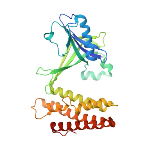
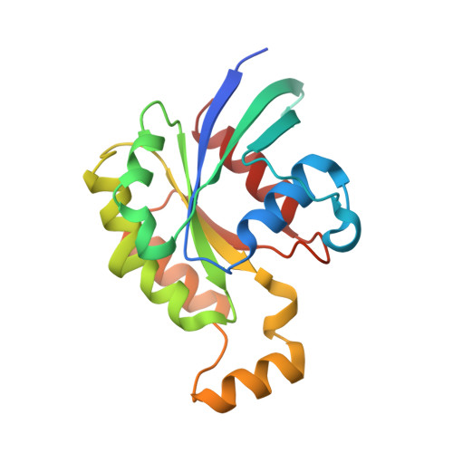
    Dedicator of cytokinesis 10 (DOCK10), an evolutionarily conserved guanine nucleotide exchange factor (GEF) for Rho GTPases, has the unique specificity within the DOCK-D subfamily to activate both Cdc42 and Rac, but the structural bases for these activities remained unknown. Here we present the crystal structures of the catalytic DHR2 domain of mouse DOCK10, complexed with either Cdc42 or Rac1. The structures revealed that DOCK10 DHR2 binds to Cdc42 or Rac1 by slightly changing the arrangement of its two catalytic lobes. DOCK10 also has a flexible binding pocket for the 56th GTPase residue, allowing a novel interaction with Trp56 Rac1 . The conserved residues in switch 1 of Cdc42 and Rac1 showed common interactions with the unique Lys-His sequence in the β5/β6 loop of DOCK10 DHR2 . However, the interaction of switch 1 in Rac1 was less stable than that of switch 1 in Cdc42, due to amino acid differences at positions 27 and 30. Structure-based mutagenesis identified the DOCK10 residues that determine the Cdc42/Rac1 dual specificity.


  • Organizational Affiliation

    Laboratory for Protein Functional and Structural Biology, RIKEN Center for Biosystems Dynamics Research, 1-7-22 Suehiro-cho, Tsurumi-ku, Yokohama, Kanagawa, 230-0045, Japan. Electronic address: kukimoto@riken.jp.


Macromolecules
Find similar proteins by:  (by identity cutoff)  |  3D Structure
Entity ID: 1
MoleculeChains Sequence LengthOrganismDetailsImage
Dedicator of cytokinesis protein 10282Mus musculusMutation(s): 0 
Gene Names: Dock10Kiaa0694Ziz3
UniProt & NIH Common Fund Data Resources
Find proteins for Q8BZN6 (Mus musculus)
Explore Q8BZN6 
Go to UniProtKB:  Q8BZN6
IMPC:  MGI:2146320
Entity Groups  
Sequence Clusters30% Identity50% Identity70% Identity90% Identity95% Identity100% Identity
UniProt GroupQ8BZN6
Sequence Annotations
Expand
  • Reference Sequence
Find similar proteins by:  (by identity cutoff)  |  3D Structure
Entity ID: 2
MoleculeChains Sequence LengthOrganismDetailsImage
Ras-related C3 botulinum toxin substrate 1184Homo sapiensMutation(s): 0 
Gene Names: RAC1TC25MIG5
EC: 3.6.5.2
UniProt & NIH Common Fund Data Resources
Find proteins for P63000 (Homo sapiens)
Explore P63000 
Go to UniProtKB:  P63000
PHAROS:  P63000
GTEx:  ENSG00000136238 
Entity Groups  
Sequence Clusters30% Identity50% Identity70% Identity90% Identity95% Identity100% Identity
UniProt GroupP63000
Sequence Annotations
Expand
  • Reference Sequence
Small Molecules
Ligands 1 Unique
IDChains Name / Formula / InChI Key2D Diagram3D Interactions
SO4
Query on SO4

Download Ideal Coordinates CCD File 
C [auth B]SULFATE ION
O4 S
QAOWNCQODCNURD-UHFFFAOYSA-L
Experimental Data & Validation

Experimental Data

  • Method: X-RAY DIFFRACTION
  • Resolution: 2.43 Å
  • R-Value Free: 0.280 
  • R-Value Work: 0.220 
  • R-Value Observed: 0.225 
  • Space Group: C 2 2 21
Unit Cell:
Length ( Å )Angle ( ˚ )
a = 60.28α = 90
b = 106.86β = 90
c = 187.11γ = 90
Software Package:
Software NamePurpose
PHENIXrefinement
XDSdata reduction
XDSdata scaling
PHASERphasing

Structure Validation

View Full Validation Report


Entry History & Funding Information

Deposition Data


Funding OrganizationLocationGrant Number
Japan Society for the Promotion of Science (JSPS)Japan--

Revision History  (Full details and data files)

  • Version 1.0: 2023-03-15
    Type: Initial release