Unusual photodynamic characteristics of the light-oxygen-voltage domain of phototropin linked to terrestrial adaptation of Klebsormidium nitens.
Sharma, S., Gautam, A.K., Singh, R., Gourinath, S., Kateriya, S.(2024) FEBS J 291: 5156-5176
- PubMed: 39344087 Search on PubMed
- DOI: https://doi.org/10.1111/febs.17284
- Primary Citation Related Structures:
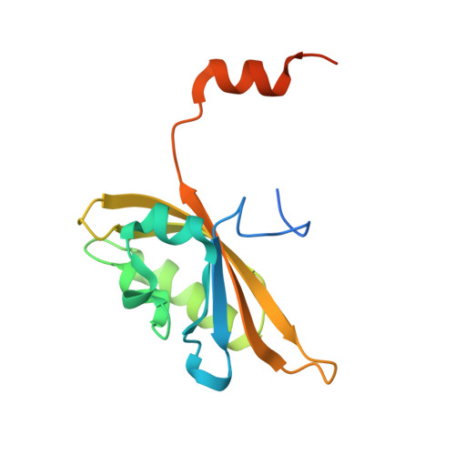
8I11, 8IL9, 8IYN, 8J68 - PubMed Abstract:
Phototropin (Phot), a blue light-sensing LOV domain protein, mediates blue light responses and is evolutionarily conserved across the green lineage. Klebsormidium nitens, a green terrestrial alga, presents a valuable opportunity to study adaptive responses from aquatic to land habitat transitions. We determined the crystal structure of Klebsormidium nitens Phot LOV1 domain (KnLOV1) in the dark and engineered different mutations (R60K, Q122N, and D33N) to modulate the lifetime of the photorecovery cycle. We observed unusual, slow recovery kinetics in the wild-type KnLOV1 domain (τ = 41 ± 3 min) compared to different mutants (R60K: τ = 2.0 ± 0.1 min, Q122N: τ = 1.7 ± 0.1 min, D33N: τ = 9.6 ± 0.1 min). Crystal structures of wild-type KnLOV1 and mutants revealed subtle but critical changes near the protein chromophore that is responsible for modulating protein dark recovery time. Our findings shed light on the unique structural and biochemical characteristics of the newly studied KnLOV1 and its evolutionary importance for phototropin-mediated physiology.
- Laboratory of Optobiotechnology, School of Biotechnology, Jawaharlal Nehru University, New Delhi, India.
Organizational Affiliation: