Two different alanine dehydrogenases from Geobacillus kaustophilus: Their biochemical characteristics and differential expression in vegetative cells and spores.
Maeno, M., Ohmori, T., Nukada, D., Sakuraba, H., Satomura, T., Ohshima, T.(2023) Biochim Biophys Acta Proteins Proteom 1871: 140904-140904
- PubMed: 36918121 Search on PubMed
- DOI: https://doi.org/10.1016/j.bbapap.2023.140904
- Primary Citation Related Structures:
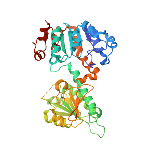
8HYE, 8HYH - PubMed Abstract:
Two putative alanine dehydrogenase (AlaDH) genes (GK2752 and GK3448) were found in the genome of a thermophilic spore-forming bacterium, Geobacillus kaustophilus. The amino acid sequences deduced from the two genes showed mutually high homology (71%), and the phylogenetic tree based on the amino acid sequences of the two putative AlaDHs and the homologous proteins showed that the two putative AlaDH genes (GK2752 and GK3448) belong to different groups. Both of the recombinant gene products exhibited high NAD + -dependent AlaDH activity and were purified to homogeneity and characterized in detail. Both enzymes showed high stability against low and high pHs and high temperatures (70 °C). Kinetic analyses showed that the activities of both enzymes proceeded according to the same sequentially ordered Bi-Ter mechanism. X-ray crystallographic analysis showed the two AlaDHs to have similar homohexameric structures. Notably, GK3448-AlaDH was detected in vegetative cells of G. kaustophilus but not spores, while GK2752-AlaDH was present only in the spores. This is the first report showing the presence of two AlaDHs separately expressed in vegetative cells and spores.
- Department of Biomedical Engineering, Osaka Institute of Technology, Osaka 535-8585, Japan.
Organizational Affiliation: