8HWJ

Bacterial STING from Epilithonimonas lactis in complex with 3'3'-c-di-AMP


Experimental Data Snapshot

  • Method: X-RAY DIFFRACTION
  • Resolution: 2.55 Å
  • R-Value Free: 0.228 
  • R-Value Work: 0.195 

wwPDB Validation   3D Report Full Report


Ligand Structure Quality Assessment 


This is version 1.1 of the entry. See complete history


Literature

Structural insights into the regulation, ligand recognition, and oligomerization of bacterial STING.

Hou, M.H.Wang, Y.C.Yang, C.S.Liao, K.F.Chang, J.W.Shih, O.Yeh, Y.Q.Sriramoju, M.K.Weng, T.W.Jeng, U.S.Hsu, S.D.Chen, Y.

(2023) Nat Commun 14: 8519-8519

  • DOI: https://doi.org/10.1038/s41467-023-44052-x
  • Primary Citation of Related Structures:  
    8HWI, 8HWJ, 8HY8, 8HY9, 8HYN

  • PubMed Abstract: 

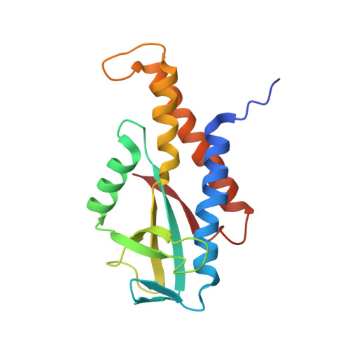
    The cyclic GMP-AMP synthase (cGAS)/stimulator of interferon gene (STING) signaling pathway plays a critical protective role against viral infections. Metazoan STING undergoes multilayers of regulation to ensure specific signal transduction. However, the mechanisms underlying the regulation of bacterial STING remain unclear. In this study, we determined the crystal structure of anti-parallel dimeric form of bacterial STING, which keeps itself in an inactive state by preventing cyclic dinucleotides access. Conformational transition between inactive and active states of bacterial STINGs provides an on-off switch for downstream signaling. Some bacterial STINGs living in extreme environment contain an insertion sequence, which we show codes for an additional long lid that covers the ligand-binding pocket. This lid helps regulate anti-phage activities. Furthermore, bacterial STING can bind cyclic di-AMP in a triangle-shaped conformation via a more compact ligand-binding pocket, forming spiral-shaped protofibrils and higher-order fibril filaments. Based on the differences between cyclic-dinucleotide recognition, oligomerization, and downstream activation of different bacterial STINGs, we proposed a model to explain structure-function evolution of bacterial STINGs.


  • Organizational Affiliation

    Genomics BioSci. & Tech. Co. Ltd., New Taipei, 221411, Taiwan.


Macromolecules
Find similar proteins by:  (by identity cutoff)  |  3D Structure
Entity ID: 1
MoleculeChains Sequence LengthOrganismDetailsImage
CD-NTase-associated protein 12
A, B, C, D
165Epilithonimonas lactisMutation(s): 0 
Gene Names: IO89_10965
UniProt
Find proteins for A0A085BE66 (Epilithonimonas lactis)
Explore A0A085BE66 
Go to UniProtKB:  A0A085BE66
Entity Groups  
Sequence Clusters30% Identity50% Identity70% Identity90% Identity95% Identity100% Identity
UniProt GroupA0A085BE66
Sequence Annotations
Expand
  • Reference Sequence
Small Molecules
Ligands 1 Unique
IDChains Name / Formula / InChI Key2D Diagram3D Interactions
2BA (Subject of Investigation/LOI)
Query on 2BA

Download Ideal Coordinates CCD File 
E [auth B],
F [auth C]
(2R,3R,3aS,5R,7aR,9R,10R,10aS,12R,14aR)-2,9-bis(6-amino-9H-purin-9-yl)octahydro-2H,7H-difuro[3,2-d:3',2'-j][1,3,7,9,2,8 ]tetraoxadiphosphacyclododecine-3,5,10,12-tetrol 5,12-dioxide
C20 H24 N10 O12 P2
PDXMFTWFFKBFIN-XPWFQUROSA-N
Experimental Data & Validation

Experimental Data

  • Method: X-RAY DIFFRACTION
  • Resolution: 2.55 Å
  • R-Value Free: 0.228 
  • R-Value Work: 0.195 
  • Space Group: C 1 2 1
Unit Cell:
Length ( Å )Angle ( ˚ )
a = 157.171α = 90
b = 52.713β = 114.883
c = 88.932γ = 90
Software Package:
Software NamePurpose
REFMACrefinement
HKL-2000data scaling
MOLREPphasing
HKL-2000data reduction

Structure Validation

View Full Validation Report


Ligand Structure Quality Assessment 


Entry History & Funding Information

Deposition Data


Funding OrganizationLocationGrant Number
Ministry of Science and Technology (MoST, Taiwan)Taiwan109-2311-B241-001
Ministry of Science and Technology (MoST, Taiwan)Taiwan111-2311-B-039-001-MY3

Revision History  (Full details and data files)

  • Version 1.0: 2024-01-03
    Type: Initial release
  • Version 1.1: 2024-01-24
    Changes: Database references