8HVQ | pdb_00008hvq

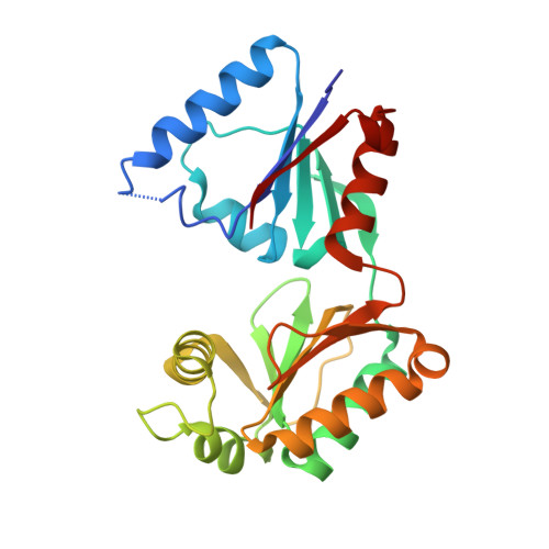
Crystal structure of haloacid dehalogenase-like hydrolase family enzyme from Staphylococcus lugdunensis


Experimental Data Snapshot

  • Method: X-RAY DIFFRACTION
  • Resolution: 1.73 Å
  • R-Value Free: 
    0.199 (Depositor), 0.203 (DCC) 
  • R-Value Work: 
    0.151 (Depositor) 
  • R-Value Observed: 
    0.153 (Depositor) 

Starting Model: in silico
View more details

wwPDB Validation 3D Report Full Report

Validation slider image for 8HVQ

This is version 1.1 of the entry. See complete history

Literature

Characterization of haloacid dehalogenase superfamily acid phosphatase from Staphylococcus lugdunensis.

Kaur, H.Rode, S.Kp, S.Mahto, J.K.Alam, M.S.Gupta, D.N.Kar, B.Singla, J.Kumar, P.Sharma, A.K.

(2024) Arch Biochem Biophys 753: 109888-109888

  • DOI: https://doi.org/10.1016/j.abb.2024.109888
  • Primary Citation Related Structures: 
    8HVQ

  • PubMed Abstract: 

    The haloacid dehalogenase superfamily implicated in bacterial pathogenesis comprises different enzymes having roles in many metabolic pathways. Staphylococcus lugdunensis, a Gram-positive bacterium, is an opportunistic human pathogen causing infections in the central nervous system, urinary tract, bones, peritoneum, systemic conditions and cutaneous infection. The haloacid dehalogenase superfamily proteins play a significant role in the pathogenicity of certain bacteria, facilitating invasion, survival, and proliferation within host cells. The genome of S. lugdunensis encodes more than ten proteins belonging to this superfamily. However, none of them have been characterized. The present work reports the characterization of one of the haloacid dehalogenase superfamily proteins (SLHAD1) from Staphylococcus lugdunensis. The functional analysis revealed that SLHAD1 is a metal-dependent acid phosphatase, which catalyzes the dephosphorylation of phosphorylated metabolites of cellular pathways, including glycolysis, gluconeogenesis, nucleotides, and thiamine metabolism. Based on the substrate specificity and genomic analysis, the physiological function of SLHAD1 in thiamine metabolism has been tentatively assigned. The crystal structure of SLHAD1, lacking 49 residues at the C-terminal, was determined at 1.7 Å resolution with a homodimer in the asymmetric unit. It was observed that SLHAD1 exhibited time-dependent cleavage at a specific point, occurring through a self-initiated process. A combination of bioinformatics, biochemical, biophysical, and structural studies explored unique features of SLHAD1. Overall, the study revealed a detailed characterization of a critical enzyme of the human pathogen Staphylococcus lugdunensis, associated with several life-threatening infections.


  • Organizational Affiliation
    • Department of Biosciences and Bioengineering, Indian Institute of Technology Roorkee, Roorkee-247667, India.

Macromolecule Content 

  • Total Structure Weight: 53.13 kDa 
  • Atom Count: 4,392 
  • Modeled Residue Count: 460 
  • Deposited Residue Count: 466 
  • Unique protein chains: 1

Macromolecules

Find similar proteins by:|  3D Structure
Entity ID: 1
MoleculeChains  Sequence LengthOrganismDetailsImage
Cof-type HAD-IIB family hydrolase
A, B
233Staphylococcus lugdunensisMutation(s): 0 
Gene Names: EQ812_11540
UniProt
Find proteins for A0A292DKY6 (Staphylococcus lugdunensis)
Explore A0A292DKY6 
Go to UniProtKB:  A0A292DKY6
Entity Groups
Sequence Clusters30% Identity50% Identity70% Identity90% Identity95% Identity100% Identity
UniProt GroupA0A292DKY6
Sequence Annotations
Expand
Reference Sequence

Small Molecules

Ligands 4 Unique
IDChains Name / Formula / InChI Key2D Diagram3D Interactions
PEG

Query on PEG


Download:Ideal Coordinates CCD File
C [auth A]DI(HYDROXYETHYL)ETHER
C4 H10 O3
MTHSVFCYNBDYFN-UHFFFAOYSA-N
OXM

Query on OXM


Download:Ideal Coordinates CCD File
E [auth A],
I [auth B]
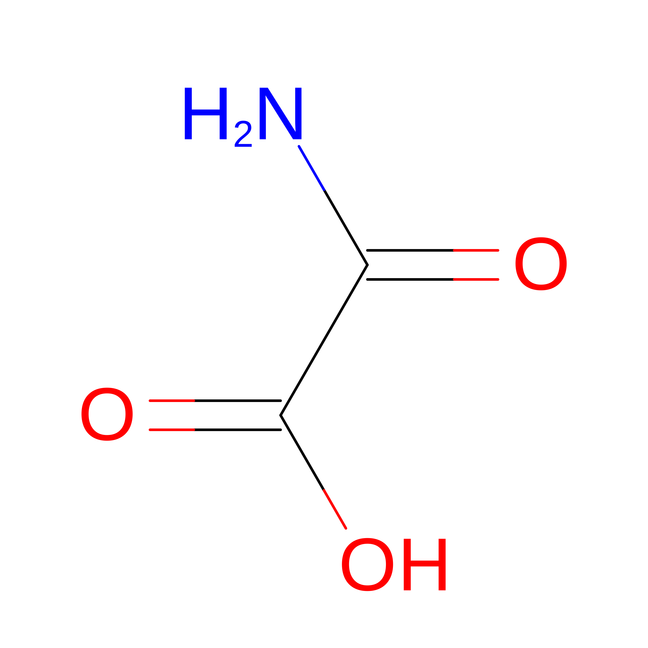
OXAMIC ACID
C2 H3 N O3
SOWBFZRMHSNYGE-UHFFFAOYSA-N
EDO

Query on EDO


Download:Ideal Coordinates CCD File
D [auth A],
G [auth A],
H [auth B],
J [auth B],
K [auth B]
1,2-ETHANEDIOL
C2 H6 O2
LYCAIKOWRPUZTN-UHFFFAOYSA-N
FMT

Query on FMT


Download:Ideal Coordinates CCD File
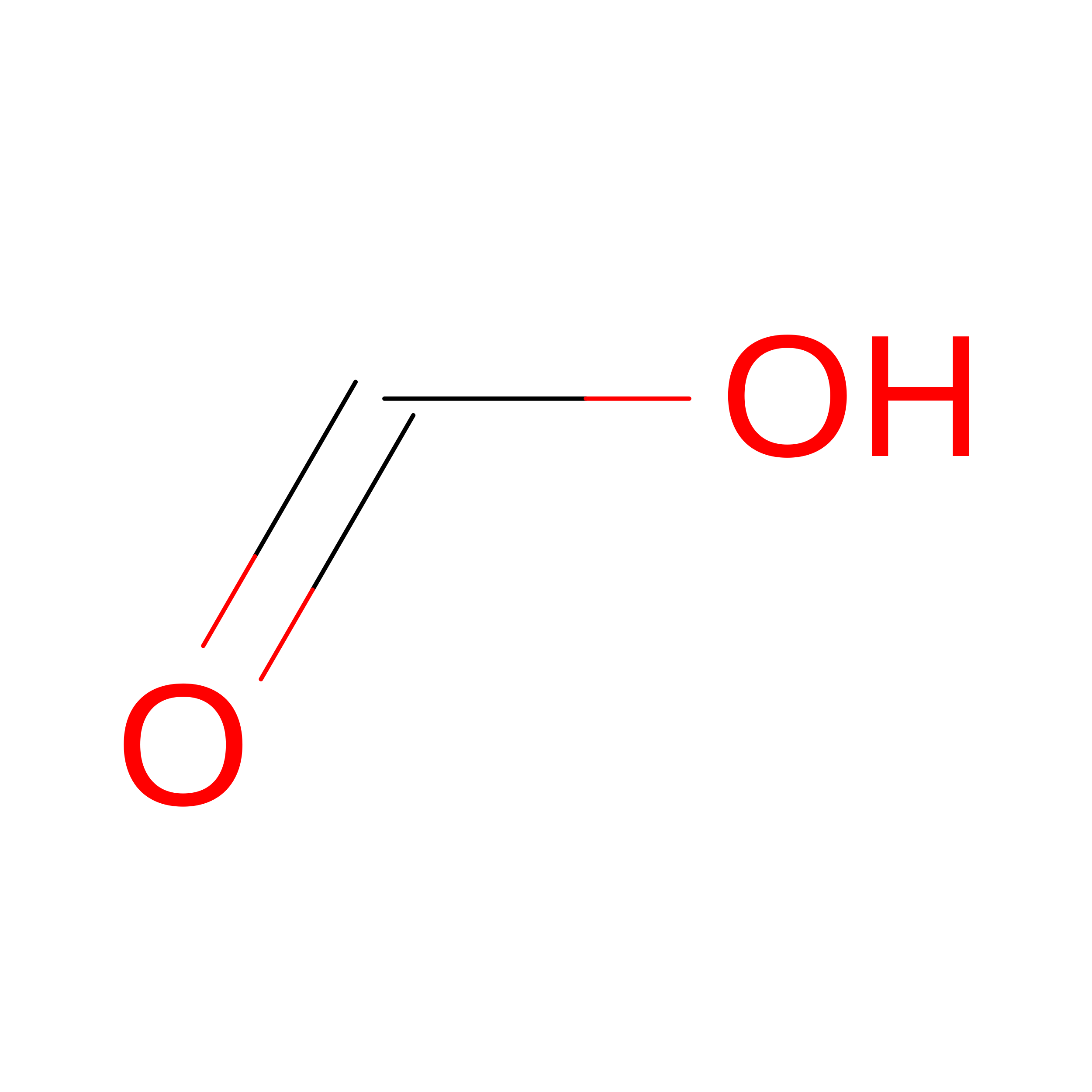
F [auth A]FORMIC ACID
C H2 O2
BDAGIHXWWSANSR-UHFFFAOYSA-N

Experimental Data & Validation

Experimental Data

  • Method: X-RAY DIFFRACTION
  • Resolution: 1.73 Å
  • R-Value Free:  0.199 (Depositor), 0.203 (DCC) 
  • R-Value Work:  0.151 (Depositor) 
  • R-Value Observed: 0.153 (Depositor) 
Space Group: P 21 21 21
Unit Cell:
Length ( Å )Angle ( ˚ )
a = 36.947α = 90
b = 121.387β = 90
c = 134.405γ = 90
Software Package:
Software NamePurpose
REFMACrefinement
autoPROCdata reduction
Aimlessdata scaling
MOLREPphasing

Structure Validation

View Full Validation Report


Entry History 

& Funding Information

Deposition Data


Funding OrganizationLocationGrant Number
Department of Science & Technology (DST, India)India--

Revision History  (Full details and data files)

  • Version 1.0: 2023-12-27
    Type: Initial release
  • Version 1.1: 2024-05-29
    Changes: Database references