8HO1

Crystal structure of cytochrome P450 NasF5053 mutant F387G


Experimental Data Snapshot

  • Method: X-RAY DIFFRACTION
  • Resolution: 2.00 Å
  • R-Value Free: 0.216 
  • R-Value Work: 0.172 
  • R-Value Observed: 0.174 

wwPDB Validation   3D Report Full Report


Ligand Structure Quality Assessment 


This is version 1.4 of the entry. See complete history


Literature

Engineering the Substrate Specificity of a P450 Dimerase Enables the Collective Biosynthesis of Heterodimeric Tryptophan-Containing Diketopiperazines.

Sun, C.Ma, B.D.Li, G.Tian, W.Yang, L.Peng, H.Lin, Z.Deng, Z.Kong, X.D.Qu, X.

(2023) Angew Chem Int Ed Engl 62: e202304994-e202304994

  • DOI: https://doi.org/10.1002/anie.202304994
  • Primary Citation of Related Structures:  
    8HNY, 8HNZ, 8HO0, 8HO1

  • PubMed Abstract: 

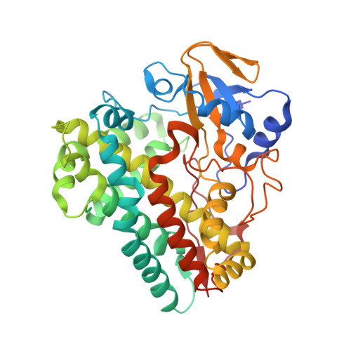
    Heterodimeric tryptophan-containing diketopiperazines (HTDKPs) are an important class of bioactive secondary metabolites. Biosynthesis offers a practical opportunity to access their bioactive structural diversity, however, it is restricted by the limited substrate scopes of the HTDKPs-forming P450 dimerases. Herein, by genome mining and investigation of the sequence-product relationships, we unveiled three important residues (F387, F388 and E73) in these P450s that are pivotal for selecting different diketopiperazine (DKP) substrates in the upper binding pocket. Engineering these residues in Nas F5053 significantly expanded its substrate specificity and enabled the collective biosynthesis, including 12 self-dimerized and at least 81 cross-dimerized HTDKPs. Structural and molecular dynamics analysis of F387G and E73S revealed that they control the substrate specificity via reducing steric hindrance and regulating substrate tunnels, respectively.


  • Organizational Affiliation

    State Key Laboratory of Microbial Metabolism and School of Life Sciences and Biotechnology, Shanghai Jiao Tong University, 800 Dongchuan Rd., 200240, Shanghai, China.


Macromolecules
Find similar proteins by:  (by identity cutoff)  |  3D Structure
Entity ID: 1
MoleculeChains Sequence LengthOrganismDetailsImage
Cytochrome P450-F5053398Streptomyces sp. NRRL F-5053Mutation(s): 1 
UniProt
Find proteins for A0A8I3B027 (Streptomyces sp. NRRL F-5053)
Explore A0A8I3B027 
Go to UniProtKB:  A0A8I3B027
Entity Groups  
Sequence Clusters30% Identity50% Identity70% Identity90% Identity95% Identity100% Identity
UniProt GroupA0A8I3B027
Sequence Annotations
Expand
  • Reference Sequence
Small Molecules
Ligands 2 Unique
IDChains Name / Formula / InChI Key2D Diagram3D Interactions
HEM
Query on HEM

Download Ideal Coordinates CCD File 
B [auth A]PROTOPORPHYRIN IX CONTAINING FE
C34 H32 Fe N4 O4
KABFMIBPWCXCRK-RGGAHWMASA-L
CA
Query on CA

Download Ideal Coordinates CCD File 
C [auth A]CALCIUM ION
Ca
BHPQYMZQTOCNFJ-UHFFFAOYSA-N
Experimental Data & Validation

Experimental Data

  • Method: X-RAY DIFFRACTION
  • Resolution: 2.00 Å
  • R-Value Free: 0.216 
  • R-Value Work: 0.172 
  • R-Value Observed: 0.174 
  • Space Group: P 21 21 21
Unit Cell:
Length ( Å )Angle ( ˚ )
a = 42.107α = 90
b = 90.579β = 90
c = 95.06γ = 90
Software Package:
Software NamePurpose
PHENIXrefinement
XDSdata scaling
XDSdata reduction
PHASERphasing

Structure Validation

View Full Validation Report


Ligand Structure Quality Assessment 


Entry History & Funding Information

Deposition Data


Funding OrganizationLocationGrant Number
Ministry of Science and Technology (MoST, China)China2020YFA0907700

Revision History  (Full details and data files)

  • Version 1.0: 2023-04-19
    Type: Initial release
  • Version 1.1: 2023-05-17
    Changes: Database references
  • Version 1.2: 2023-06-28
    Changes: Database references
  • Version 1.3: 2023-07-12
    Changes: Data collection, Database references
  • Version 1.4: 2023-08-30
    Changes: Data collection