8HNJ | pdb_00008hnj

Domain-stabilized glutamine-binding protein


Experimental Data Snapshot

  • Method: X-RAY DIFFRACTION
  • Resolution: 2.03 Å
  • R-Value Free: 
    0.264 (Depositor), 0.264 (DCC) 
  • R-Value Work: 
    0.226 (Depositor), 0.227 (DCC) 
  • R-Value Observed: 
    0.228 (Depositor) 

Starting Model: in silico
View more details

wwPDB Validation 3D Report Full Report

Validation slider image for 8HNJ

Ligand Structure Quality Assessment 


This is version 1.1 of the entry. See complete history

Literature

Domain-wise dissection of thermal stability enhancement in multidomain proteins.

Oh, J.Durai, P.Kannan, P.Park, J.Yeon, Y.J.Lee, W.K.Park, K.Seo, M.H.

(2023) Int J Biol Macromol 237: 124141-124141

  • DOI: https://doi.org/10.1016/j.ijbiomac.2023.124141
  • Primary Citation Related Structures: 
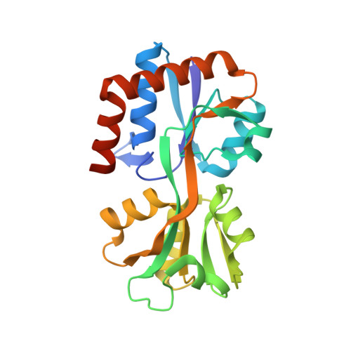
    8HNJ

  • PubMed Abstract: 

    Stability is critical for the proper functioning of all proteins. Optimization of protein thermostability is a key step in the development of industrial enzymes and biologics. Herein, we demonstrate that multidomain proteins can be stabilized significantly using domain-based engineering followed by the recombination of the optimized domains. Domain-level analysis of designed protein variants with similar structures but different thermal profiles showed that the independent enhancement of the thermostability of a constituent domain improves the overall stability of the whole multidomain protein. The crystal structure and AlphaFold-predicted model of the designed proteins via domain-recombination provided a molecular explanation for domain-based stepwise stabilization. Our study suggests that domain-based modular engineering can minimize the sequence space for calculations in computational design and experimental errors, thereby offering useful guidance for multidomain protein engineering.


  • Organizational Affiliation
    • Natural Product Research Center, Korea Institute of Science and Technology, Gangneung 25451, South Korea; Department of Biochemical Engineering, Gangneung-Wonju National University, Gangneung 25457, South Korea.

Macromolecule Content 

  • Total Structure Weight: 154.13 kDa 
  • Atom Count: 10,642 
  • Modeled Residue Count: 1,314 
  • Deposited Residue Count: 1,380 
  • Unique protein chains: 1

Macromolecules

Find similar proteins by:|  3D Structure
Entity ID: 1
MoleculeChains  Sequence LengthOrganismDetailsImage
Glutamine ABC transporter, periplasmic glutamine-binding protein GlnH
A, B, C, D, E
A, B, C, D, E, F
230Escherichia coli 908519Mutation(s): 0 
Gene Names: HMPREF1604_05361
UniProt
Find proteins for P0AEQ3 (Escherichia coli (strain K12))
Explore P0AEQ3 
Go to UniProtKB:  P0AEQ3
Entity Groups
Sequence Clusters30% Identity50% Identity70% Identity90% Identity95% Identity100% Identity
UniProt GroupP0AEQ3
Sequence Annotations
Expand
Reference Sequence

Experimental Data & Validation

Experimental Data

  • Method: X-RAY DIFFRACTION
  • Resolution: 2.03 Å
  • R-Value Free:  0.264 (Depositor), 0.264 (DCC) 
  • R-Value Work:  0.226 (Depositor), 0.227 (DCC) 
  • R-Value Observed: 0.228 (Depositor) 
Space Group: C 1 2 1
Unit Cell:
Length ( Å )Angle ( ˚ )
a = 235.1α = 90
b = 69.59β = 90.711
c = 83.74γ = 90
Software Package:
Software NamePurpose
PHENIXrefinement
autoPROCdata scaling
PHENIXphasing
HKL-2000data reduction

Structure Validation

View Full Validation Report


Ligand Structure Quality Assessment 


Entry History 

& Funding Information

Deposition Data


Funding OrganizationLocationGrant Number
Other governmentKorea, Republic Of--

Revision History  (Full details and data files)

  • Version 1.0: 2023-07-19
    Type: Initial release
  • Version 1.1: 2024-05-29
    Changes: Data collection