The Mycobacterium tuberculosis methyltransferase Rv2067c manipulates host epigenetic programming to promote its own survival.
Singh, P.R., Dadireddy, V., Udupa, S., Kalladi, S.M., Shee, S., Khosla, S., Rajmani, R.S., Singh, A., Ramakumar, S., Nagaraja, V.(2023) Nat Commun 14: 8497-8497
- PubMed: 38129415 Search on PubMedSearch on PubMed Central
- DOI: https://doi.org/10.1038/s41467-023-43940-6
- Primary Citation Related Structures:
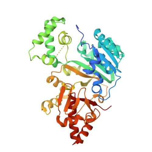
8HKR - PubMed Abstract:
Mycobacterium tuberculosis has evolved several mechanisms to counter host defense arsenals for its proliferation. Here we report that M. tuberculosis employs a multi-pronged approach to modify host epigenetic machinery for its survival. It secretes methyltransferase (MTase) Rv2067c into macrophages, trimethylating histone H3K79 in a non-nucleosomal context. Rv2067c downregulates host MTase DOT1L, decreasing DOT1L-mediated nucleosomally added H3K79me3 mark on pro-inflammatory response genes. Consequent inhibition of caspase-8-dependent apoptosis and enhancement of RIPK3-mediated necrosis results in increased pathogenesis. In parallel, Rv2067c enhances the expression of SESTRIN3, NLRC3, and TMTC1, enabling the pathogen to overcome host inflammatory and oxidative responses. We provide the structural basis for differential methylation of H3K79 by Rv2067c and DOT1L. The structures of Rv2067c and DOT1L explain how their action on H3K79 is spatially and temporally separated, enabling Rv2067c to effectively intercept the host epigenetic circuit and downstream signaling.
- Department of Microbiology & Cell Biology, Indian Institute of Science (IISc), Bengaluru, India.
Organizational Affiliation: