8HFP | pdb_00008hfp

Crystal structure of the methyl-CpG-binding domain of SETDB2 in complex with the cysteine-rich domain of C11orf46 protein


Experimental Data Snapshot

  • Method: X-RAY DIFFRACTION
  • Resolution: 1.82 Å
  • R-Value Free: 
    0.227 (Depositor), 0.235 (DCC) 
  • R-Value Work: 
    0.186 (Depositor), 0.194 (DCC) 
  • R-Value Observed: 
    0.188 (Depositor) 

Starting Model: experimental
View more details

wwPDB Validation 3D Report Full Report

Validation slider image for 8HFP

This is version 1.2 of the entry. See complete history

Literature

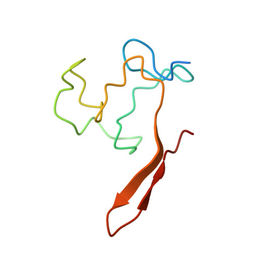
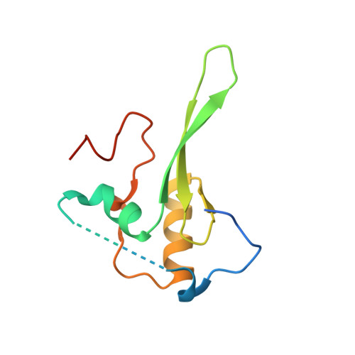
Structural evidence for protein-protein interaction between the non-canonical methyl-CpG-binding domain of SETDB proteins and C11orf46.

Mahana, Y.Ariyoshi, M.Nozawa, R.S.Shibata, S.Nagao, K.Obuse, C.Shirakawa, M.

(2024) Structure 32: 304-315.e5

  • DOI: https://doi.org/10.1016/j.str.2023.12.001
  • Primary Citation Related Structures: 
    8HFP

  • PubMed Abstract: 

    SETDB1 and SETDB2 mediate trimethylation of histone H3 lysine 9 (H3K9), an epigenetic hallmark of repressive chromatin. They contain a non-canonical methyl-CpG-binding domain (MBD) and bifurcated SET domain, implying interplay between H3K9 trimethylation and DNA methylation in SETDB functions. Here, we report the crystal structure of human SETDB2 MBD bound to the cysteine-rich domain of a zinc-binding protein, C11orf46. SETDB2 MBD comprises the conserved MBD core and a unique N-terminal extension. Although the MBD core has the conserved basic concave surface for DNA binding, it utilizes it for recognition of the cysteine-rich domain of C11orf46. This interaction involves the conserved arginine finger motif and the unique N-terminal extension of SETDB2 MBD, with a contribution from intermolecular β-sheet formation. Thus, the non-canonical MBD of SETDB1/2 seems to have lost methylated DNA-binding ability but gained a protein-protein interaction surface. Our findings provide insight into the molecular assembly of SETDB-associated repression complexes.


  • Organizational Affiliation
    • Department of Molecular Engineering, Graduate School of Engineering, Kyoto University, Nishikyo-Ku, Kyoto 615-8510, Japan.

Macromolecule Content 

  • Total Structure Weight: 42.93 kDa 
  • Atom Count: 2,568 
  • Modeled Residue Count: 306 
  • Deposited Residue Count: 370 
  • Unique protein chains: 2

Macromolecules

Find similar proteins by:|  3D Structure
Entity ID: 1
MoleculeChains  Sequence LengthOrganismDetailsImage
ARL14 effector proteinA,
C [auth B]
77Homo sapiensMutation(s): 0 
Gene Names: ARL14EPARF7EPC11orf46
UniProt & NIH Common Fund Data Resources
Find proteins for Q8N8R7 (Homo sapiens)
Explore Q8N8R7 
Go to UniProtKB:  Q8N8R7
PHAROS:  Q8N8R7
GTEx:  ENSG00000152219 
Entity Groups
Sequence Clusters30% Identity50% Identity70% Identity90% Identity95% Identity100% Identity
UniProt GroupQ8N8R7
Sequence Annotations
Expand
Reference Sequence
Find similar proteins by:|  3D Structure
Entity ID: 2
MoleculeChains  Sequence LengthOrganismDetailsImage
Histone-lysine N-methyltransferase SETDB2B [auth C],
D
108Homo sapiensMutation(s): 0 
Gene Names: SETDB2C13orf4CLLD8KMT1F
EC: 2.1.1.366
UniProt & NIH Common Fund Data Resources
Find proteins for Q96T68 (Homo sapiens)
Explore Q96T68 
Go to UniProtKB:  Q96T68
PHAROS:  Q96T68
GTEx:  ENSG00000136169 
Entity Groups
Sequence Clusters30% Identity50% Identity70% Identity90% Identity95% Identity100% Identity
UniProt GroupQ96T68
Sequence Annotations
Expand
Reference Sequence

Small Molecules

Ligands 2 Unique
IDChains Name / Formula / InChI Key2D Diagram3D Interactions
SO4

Query on SO4


Download:Ideal Coordinates CCD File
G [auth A]
I [auth C]
J [auth C]
M [auth B]
N [auth B]
G [auth A],
I [auth C],
J [auth C],
M [auth B],
N [auth B],
P [auth D]
SULFATE ION
O4 S
QAOWNCQODCNURD-UHFFFAOYSA-L
ZN

Query on ZN


Download:Ideal Coordinates CCD File
E [auth A]
F [auth A]
H [auth C]
K [auth B]
L [auth B]
E [auth A],
F [auth A],
H [auth C],
K [auth B],
L [auth B],
O [auth D]
ZINC ION
Zn
PTFCDOFLOPIGGS-UHFFFAOYSA-N

Experimental Data & Validation

Experimental Data

  • Method: X-RAY DIFFRACTION
  • Resolution: 1.82 Å
  • R-Value Free:  0.227 (Depositor), 0.235 (DCC) 
  • R-Value Work:  0.186 (Depositor), 0.194 (DCC) 
  • R-Value Observed: 0.188 (Depositor) 
Space Group: P 31 2 1
Unit Cell:
Length ( Å )Angle ( ˚ )
a = 69.97α = 90
b = 69.97β = 90
c = 116.88γ = 120
Software Package:
Software NamePurpose
REFMACrefinement
Cootmodel building
BUCCANEERmodel building
SHELXCDphasing
Aimlessdata scaling
XDSdata reduction

Structure Validation

View Full Validation Report


Entry History 

& Funding Information

Deposition Data


Funding OrganizationLocationGrant Number
Japan Society for the Promotion of Science (JSPS)Japan21K06048

Revision History  (Full details and data files)

  • Version 1.0: 2023-11-22
    Type: Initial release
  • Version 1.1: 2024-06-26
    Changes: Database references
  • Version 1.2: 2026-03-04
    Changes: Refinement description, Structure summary