8HE3

Crystal structure of importin-alpha1 bound to the HIF-1alpha nuclear localization signal (delta 724-751)


Experimental Data Snapshot

  • Method: X-RAY DIFFRACTION
  • Resolution: 1.90 Å
  • R-Value Free: 0.207 
  • R-Value Work: 0.189 
  • R-Value Observed: 0.190 

wwPDB Validation   3D Report Full Report


This is version 1.2 of the entry. See complete history


Literature

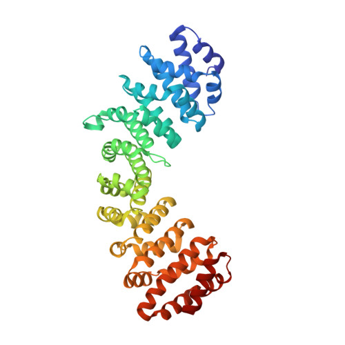
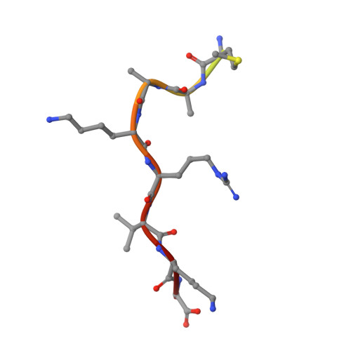
Structures of importin-alpha bound to the wild-type and an internal deletion mutant of the bipartite nuclear localization signal of HIF-1 alpha.

Matsuura, Y.Miyawaki, K.

(2023) Biochem Biophys Res Commun 652: 1-5

  • DOI: https://doi.org/10.1016/j.bbrc.2023.02.036
  • Primary Citation of Related Structures:  
    8HE0, 8HE3

  • PubMed Abstract: 

    Hypoxia-inducible factor 1 (HIF-1) is a heterodimeric transcription factor that plays an important role as a master regulator of oxygen homeostasis. The activity of HIF-1 is regulated in part by dynamic intracellular trafficking of its α subunit (HIF-1α) that can shuttle between the nucleus and cytoplasm. It has been shown that nuclear localization of HIF-1α requires a variant of classic nuclear localization signal (NLS) and that an internal deletion of the amino acid residues (residues 724-751) in the NLS almost abolish the nuclear localization. Here we report the X-ray crystal structure of the nuclear import adaptor importin-α1 bound to the wild-type HIF-1α NLS at 1.8 Å resolution and of importin-α1 bound to the Δ724-751 mutant of the HIF-1α NLS at 1.9 Å resolution. In the wild-type structure, two basic clusters in the HIF-1α NLS made extensive interactions with importin-α1 on two sites (the major site and the minor site). In the mutant structure, the NLS residues still interacted extensively with the major site on importin-α1, but the interactions with the minor site were not observed. The structural data, together with computational analyses of binding free energies, indicate that the loss of the minor-site interactions inhibit nuclear accumulation of HIF-1α.


  • Organizational Affiliation

    Department of Pharmaceutical Sciences, School of Pharmacy, International University of Health and Welfare, Japan; Division of Biological Science, Graduate School of Science, Nagoya University, Japan. Electronic address: ymatsuura@iuhw.ac.jp.


Macromolecules
Find similar proteins by:  (by identity cutoff)  |  3D Structure
Entity ID: 1
MoleculeChains Sequence LengthOrganismDetailsImage
Importin subunit alpha-1428Mus musculusMutation(s): 0 
Gene Names: Kpna2Rch1
UniProt & NIH Common Fund Data Resources
Find proteins for P52293 (Mus musculus)
Explore P52293 
Go to UniProtKB:  P52293
IMPC:  MGI:103561
Entity Groups  
Sequence Clusters30% Identity50% Identity70% Identity90% Identity95% Identity100% Identity
UniProt GroupP52293
Sequence Annotations
Expand
  • Reference Sequence

Find similar proteins by:  Sequence   |   3D Structure  

Entity ID: 2
MoleculeChains Sequence LengthOrganismDetailsImage
Hypoxia-inducible factor 1-alpha15Homo sapiensMutation(s): 0 
Gene Names: HIF1A
UniProt & NIH Common Fund Data Resources
Find proteins for Q16665 (Homo sapiens)
Explore Q16665 
Go to UniProtKB:  Q16665
PHAROS:  Q16665
GTEx:  ENSG00000100644 
Entity Groups  
Sequence Clusters30% Identity50% Identity70% Identity90% Identity95% Identity100% Identity
UniProt GroupQ16665
Sequence Annotations
Expand
  • Reference Sequence
Experimental Data & Validation

Experimental Data

  • Method: X-RAY DIFFRACTION
  • Resolution: 1.90 Å
  • R-Value Free: 0.207 
  • R-Value Work: 0.189 
  • R-Value Observed: 0.190 
  • Space Group: P 21 21 21
Unit Cell:
Length ( Å )Angle ( ˚ )
a = 78.82α = 90
b = 89.65β = 90
c = 100.17γ = 90
Software Package:
Software NamePurpose
PHENIXrefinement
MOSFLMdata reduction
SCALEITdata scaling
MOLREPphasing

Structure Validation

View Full Validation Report


Entry History & Funding Information

Deposition Data

  • Released Date: 2023-02-22 
  • Deposition Author(s): Matsuura, Y.

Funding OrganizationLocationGrant Number
Japan Society for the Promotion of Science (JSPS)Japan19K06511

Revision History  (Full details and data files)

  • Version 1.0: 2023-02-22
    Type: Initial release
  • Version 1.1: 2023-03-08
    Changes: Database references
  • Version 1.2: 2023-11-29
    Changes: Data collection, Refinement description