8HC0

Crystal structure of the extracellular domains of GPR110


Experimental Data Snapshot

  • Method: X-RAY DIFFRACTION
  • Resolution: 2.90 Å
  • R-Value Free: 0.292 
  • R-Value Work: 0.233 
  • R-Value Observed: 0.235 

wwPDB Validation   3D Report Full Report


Ligand Structure Quality Assessment 


This is version 1.2 of the entry. See complete history


Literature

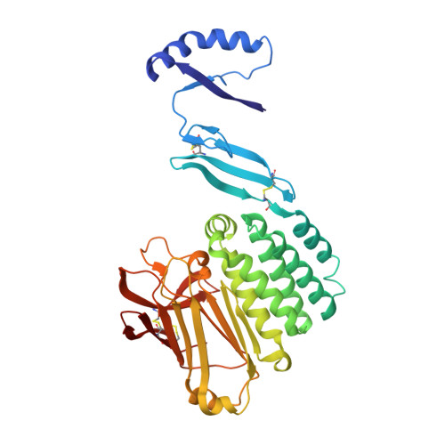
Crystal Structure of the Extracellular Domains of GPR110.

Wang, F.Wang, Y.Qiu, W.Zhang, Q.Yang, H.Song, G.

(2023) J Mol Biol 435: 167979-167979

  • DOI: https://doi.org/10.1016/j.jmb.2023.167979
  • Primary Citation of Related Structures:  
    8HC0

  • PubMed Abstract: 

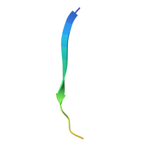
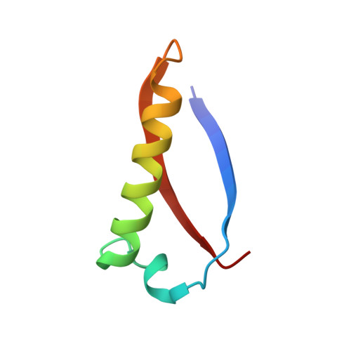
    Adhesion G protein-coupled receptors (aGPCRs) play a pivotal role in human immune responses, cellular communication, organ development, and other processes. GPR110 belongs to the aGPCR subfamily VI and was initially identified as an oncogene involved in lung and prostate cancers. GPR110 contains tandem adhesion domains at the extracellular region that mediate inter-cellular signaling. However, the structural organization and signaling mechanism for these tandem domains remain unclear. Here, we report the crystal structure of a GPR110 fragment composing the SEA, HormR, and GAIN domains at 2.9 Å resolution. The structure together with MD simulations reveal rigid connections between these domains that are stabilized by complementary interfaces. Strikingly, we found N-linked carbohydrates attached to N389 of the GAIN domain form extensive contacts with the preceding HormR domain. These interactions appear to be critical for folding, as removal of the glycosylation site greatly decreases expression of the GPR110 extracellular fragment. We further demonstrate that the ligand synaptamide fits well within the hydrophobic pocket occupied by the Stachel peptide in the rest state. This suggests that the agonist may function by removing the Stachel peptide which in turn redocks to the orthosteric pocket for receptor activation. Taken together, our structural findings and analyses provide novel insights into the activation mechanism for aGPCRs.


  • Organizational Affiliation

    Shanghai Key Laboratory of Regulatory Biology, Institute of Biomedical Sciences and School of Life Sciences, East China Normal University, Shanghai 200241, China.


Macromolecules
Find similar proteins by:  (by identity cutoff)  |  3D Structure
Entity ID: 1
MoleculeChains Sequence LengthOrganismDetailsImage
Adhesion G-protein coupled receptor F1360Homo sapiensMutation(s): 0 
Gene Names: ADGRF1GPR110PGR19
UniProt & NIH Common Fund Data Resources
Find proteins for Q5T601 (Homo sapiens)
Explore Q5T601 
Go to UniProtKB:  Q5T601
PHAROS:  Q5T601
GTEx:  ENSG00000153292 
Entity Groups  
Sequence Clusters30% Identity50% Identity70% Identity90% Identity95% Identity100% Identity
UniProt GroupQ5T601
Sequence Annotations
Expand
  • Reference Sequence

Find similar proteins by:  Sequence   |   3D Structure  

Entity ID: 2
MoleculeChains Sequence LengthOrganismDetailsImage
Adhesion G-protein coupled receptor F1B [auth C]21Homo sapiensMutation(s): 0 
Gene Names: ADGRF1GPR110PGR19
UniProt & NIH Common Fund Data Resources
Find proteins for Q5T601 (Homo sapiens)
Explore Q5T601 
Go to UniProtKB:  Q5T601
PHAROS:  Q5T601
GTEx:  ENSG00000153292 
Entity Groups  
Sequence Clusters30% Identity50% Identity70% Identity90% Identity95% Identity100% Identity
UniProt GroupQ5T601
Sequence Annotations
Expand
  • Reference Sequence
Find similar proteins by:  (by identity cutoff)  |  3D Structure
Entity ID: 3
MoleculeChains Sequence LengthOrganismDetailsImage
Adhesion G-protein coupled receptor F1C [auth B]57Homo sapiensMutation(s): 0 
Gene Names: ADGRF1GPR110PGR19
UniProt & NIH Common Fund Data Resources
Find proteins for Q5T601 (Homo sapiens)
Explore Q5T601 
Go to UniProtKB:  Q5T601
PHAROS:  Q5T601
GTEx:  ENSG00000153292 
Entity Groups  
Sequence Clusters30% Identity50% Identity70% Identity90% Identity95% Identity100% Identity
UniProt GroupQ5T601
Sequence Annotations
Expand
  • Reference Sequence
Oligosaccharides

Help

Entity ID: 4
MoleculeChains Length2D Diagram Glycosylation3D Interactions
alpha-D-mannopyranose-(1-3)-alpha-D-mannopyranose-(1-6)-[alpha-D-mannopyranose-(1-3)]alpha-D-mannopyranose-(1-4)-2-acetamido-2-deoxy-beta-D-glucopyranose-(1-4)-2-acetamido-2-deoxy-beta-D-glucopyranose
D
6N-Glycosylation
Glycosylation Resources
GlyTouCan:  G98400NB
GlyCosmos:  G98400NB
GlyGen:  G98400NB
Experimental Data & Validation

Experimental Data

  • Method: X-RAY DIFFRACTION
  • Resolution: 2.90 Å
  • R-Value Free: 0.292 
  • R-Value Work: 0.233 
  • R-Value Observed: 0.235 
  • Space Group: C 1 2 1
Unit Cell:
Length ( Å )Angle ( ˚ )
a = 134.825α = 90
b = 47.993β = 127.35
c = 87.023γ = 90
Software Package:
Software NamePurpose
PHENIXrefinement
XDSdata scaling
PDB_EXTRACTdata extraction
XDSdata reduction
PHASERphasing

Structure Validation

View Full Validation Report


Ligand Structure Quality Assessment 


Entry History & Funding Information

Deposition Data


Funding OrganizationLocationGrant Number
National Natural Science Foundation of China (NSFC)China32171215

Revision History  (Full details and data files)

  • Version 1.0: 2023-09-13
    Type: Initial release
  • Version 1.1: 2023-09-20
    Changes: Refinement description
  • Version 1.2: 2024-05-01
    Changes: Database references