8H9P

Human ATP synthase F1 domain, state 3b


Experimental Data Snapshot

  • Method: ELECTRON MICROSCOPY
  • Resolution: 3.02 Å
  • Aggregation State: PARTICLE 
  • Reconstruction Method: SINGLE PARTICLE 

wwPDB Validation   3D Report Full Report


This is version 1.2 of the entry. See complete history


Literature

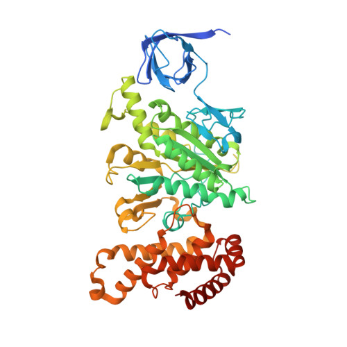
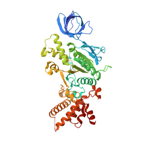
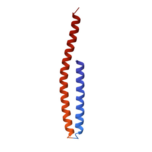
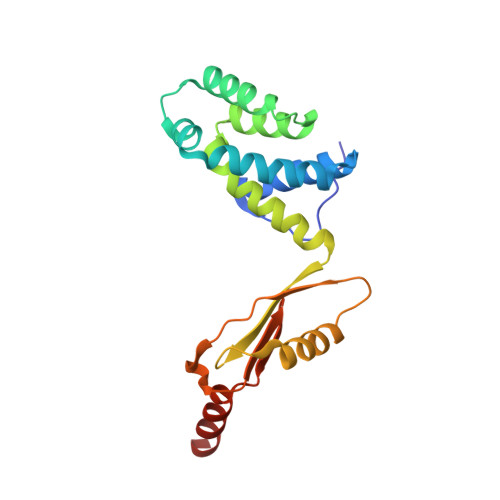
Structure of the human ATP synthase.

Lai, Y.Zhang, Y.Zhou, S.Xu, J.Du, Z.Feng, Z.Yu, L.Zhao, Z.Wang, W.Tang, Y.Yang, X.Guddat, L.W.Liu, F.Gao, Y.Rao, Z.Gong, H.

(2023) Mol Cell 83: 2137

  • DOI: https://doi.org/10.1016/j.molcel.2023.04.029
  • Primary Citation of Related Structures:  
    8H9E, 8H9F, 8H9G, 8H9I, 8H9J, 8H9K, 8H9L, 8H9M, 8H9N, 8H9P, 8H9Q, 8H9R, 8H9S, 8H9T, 8H9U, 8H9V

  • PubMed Abstract: 

    Biological energy currency ATP is produced by F 1 F o -ATP synthase. However, the molecular mechanism for human ATP synthase action remains unknown. Here, we present snapshot images for three main rotational states and one substate of human ATP synthase using cryoelectron microscopy. These structures reveal that the release of ADP occurs when the β subunit of F 1 F o -ATP synthase is in the open conformation, showing how ADP binding is coordinated during synthesis. The accommodation of the symmetry mismatch between F 1 and F o motors is resolved by the torsional flexing of the entire complex, especially the γ subunit, and the rotational substep of the c subunit. Water molecules are identified in the inlet and outlet half-channels, suggesting that the proton transfer in these two half-channels proceed via a Grotthus mechanism. Clinically relevant mutations are mapped to the structure, showing that they are mainly located at the subunit-subunit interfaces, thus causing instability of the complex.


  • Organizational Affiliation

    State Key Laboratory of Medicinal Chemical Biology and Frontiers Science Center for Cell Responses, College of Life Sciences, Nankai University, Tianjin 300071, China; Institute for Immunology, College of Life Sciences, Nankai University, Tianjin 300071, China.


Macromolecules
Find similar proteins by:  (by identity cutoff)  |  3D Structure
Entity ID: 1
MoleculeChains Sequence LengthOrganismDetailsImage
ATP synthase subunit alpha, mitochondrial
A, B, C
510Homo sapiensMutation(s): 0 
Membrane Entity: Yes 
UniProt & NIH Common Fund Data Resources
Find proteins for P25705 (Homo sapiens)
Explore P25705 
Go to UniProtKB:  P25705
PHAROS:  P25705
GTEx:  ENSG00000152234 
Entity Groups  
Sequence Clusters30% Identity50% Identity70% Identity90% Identity95% Identity100% Identity
UniProt GroupP25705
Sequence Annotations
Expand
  • Reference Sequence
Find similar proteins by:  (by identity cutoff)  |  3D Structure
Entity ID: 2
MoleculeChains Sequence LengthOrganismDetailsImage
ATP synthase subunit beta, mitochondrialD [auth E],
E [auth F],
F [auth D]
482Homo sapiensMutation(s): 0 
Membrane Entity: Yes 
UniProt & NIH Common Fund Data Resources
Find proteins for P06576 (Homo sapiens)
Explore P06576 
Go to UniProtKB:  P06576
PHAROS:  P06576
GTEx:  ENSG00000110955 
Entity Groups  
Sequence Clusters30% Identity50% Identity70% Identity90% Identity95% Identity100% Identity
UniProt GroupP06576
Sequence Annotations
Expand
  • Reference Sequence
Find similar proteins by:  (by identity cutoff)  |  3D Structure
Entity ID: 3
MoleculeChains Sequence LengthOrganismDetailsImage
ATP synthase subunit gamma, mitochondrial273Homo sapiensMutation(s): 0 
Membrane Entity: Yes 
UniProt & NIH Common Fund Data Resources
Find proteins for P36542 (Homo sapiens)
Explore P36542 
Go to UniProtKB:  P36542
PHAROS:  P36542
GTEx:  ENSG00000165629 
Entity Groups  
Sequence Clusters30% Identity50% Identity70% Identity90% Identity95% Identity100% Identity
UniProt GroupP36542
Sequence Annotations
Expand
  • Reference Sequence
Find similar proteins by:  (by identity cutoff)  |  3D Structure
Entity ID: 4
MoleculeChains Sequence LengthOrganismDetailsImage
ATP synthase subunit O, mitochondrialH [auth O]190Homo sapiensMutation(s): 0 
Membrane Entity: Yes 
UniProt & NIH Common Fund Data Resources
Find proteins for P48047 (Homo sapiens)
Explore P48047 
Go to UniProtKB:  P48047
PHAROS:  P48047
GTEx:  ENSG00000241837 
Entity Groups  
Sequence Clusters30% Identity50% Identity70% Identity90% Identity95% Identity100% Identity
UniProt GroupP48047
Sequence Annotations
Expand
  • Reference Sequence
Small Molecules
Ligands 3 Unique
IDChains Name / Formula / InChI Key2D Diagram3D Interactions
ATP (Subject of Investigation/LOI)
Query on ATP

Download Ideal Coordinates CCD File 
I [auth A],
K [auth B],
M [auth C]
ADENOSINE-5'-TRIPHOSPHATE
C10 H16 N5 O13 P3
ZKHQWZAMYRWXGA-KQYNXXCUSA-N
ADP (Subject of Investigation/LOI)
Query on ADP

Download Ideal Coordinates CCD File 
O [auth F],
Q [auth D]
ADENOSINE-5'-DIPHOSPHATE
C10 H15 N5 O10 P2
XTWYTFMLZFPYCI-KQYNXXCUSA-N
MG (Subject of Investigation/LOI)
Query on MG

Download Ideal Coordinates CCD File 
J [auth A],
L [auth B],
N [auth C],
P [auth F],
R [auth D]
MAGNESIUM ION
Mg
JLVVSXFLKOJNIY-UHFFFAOYSA-N
Experimental Data & Validation

Experimental Data

  • Method: ELECTRON MICROSCOPY
  • Resolution: 3.02 Å
  • Aggregation State: PARTICLE 
  • Reconstruction Method: SINGLE PARTICLE 

Structure Validation

View Full Validation Report


Entry History & Funding Information

Deposition Data


Funding OrganizationLocationGrant Number
National Natural Science Foundation of China (NSFC)China81520108019
National Natural Science Foundation of China (NSFC)China813300237
National Natural Science Foundation of China (NSFC)China32100976
National Natural Science Foundation of China (NSFC)China82222042

Revision History  (Full details and data files)

  • Version 1.0: 2023-05-31
    Type: Initial release
  • Version 1.1: 2023-06-07
    Changes: Database references
  • Version 1.2: 2023-07-05
    Changes: Database references