Structural characterization of the native oligomerization mode of MvaT proteins in Pseudomonas.
Vasileva, D., Suzuki-Minakuchi, C., Arakawa, T., Moriwaki, Y., Yonezawa, K., Shimizu, N., Fujimoto, Z., Terada, T., Okada, K., Nojiri, H.(2026) Microbiol Spectr : e0023526-e0023526
- PubMed: 41940658 Search on PubMed
- DOI: https://doi.org/10.1128/spectrum.00235-26
- Primary Citation Related Structures:
8H8H - PubMed Abstract:
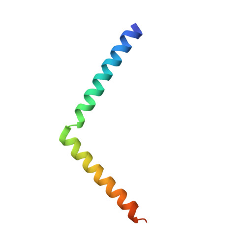
Histone-like protein H1 (H-NS) proteins are key regulators of horizontally acquired genes in enterobacteria. Functional analogs of H-NS, collectively known as H-NS family proteins, have been discovered in diverse bacteria and on large transmissible plasmids. These proteins share the ability to form oligomers through their N-terminal oligomerization domain, a feature critical for their regulatory functions. The N-terminal domain of MvaT proteins, members of the H-NS family proteins in pseudomonads, consists of a central and a terminal dimerization site. We previously elucidated the structure of the central dimerization site of TurB, an MvaT homolog in P. putida KT2440. Here, we solved the crystal and solution structures of its terminal dimerization site and generated a structural model of TurB in an oligomerized form. Our data show that the terminal dimerization site of TurB is composed of two helices connected by a flexible hinge. The results also reveal differences between the oligomerization manner of MvaT proteins and H-NS in enterobacteria. Residues forming salt bridges and hydrophobic interactions that stabilize the terminal dimerization site are relatively conserved among MvaT proteins encoded on the chromosomes of various Pseudomonas strains and plasmids. Previous studies have shown that Pseudomonas strains typically harbor plasmids encoding MvaT proteins, while enterobacteria carry plasmids encoding H-NS. Our results suggest that differences in the oligomerization domain of H-NS family proteins may be critical for the cross-talk between plasmids and the host chromosome.IMPORTANCEHorizontal gene transfer is a major driver of microbial evolution. H-NS family proteins are key factors in optimizing the transcription of newly acquired genes in host cells. Despite high sequence diversity among bacteria, these proteins share the ability to form oligomers along DNA, a critical property for their function. H-NS family proteins encoded both on large transmissible plasmids and on the chromosomes of host cells play an important role in the successful integration of new genetic sequences into the regulatory networks of microbes. Bacterial hosts typically harbor plasmids encoding the same type of H-NS family proteins as found on their chromosomes. Here, we characterized the structural basis for oligomerization of MvaT proteins, members of the H-NS family proteins in pseudomonads. Our findings reveal differences between the oligomerization mode of MvaT proteins and H-NS in enterobacteria, suggesting that these differences might impact the transmission routes of plasmids within microbial communities.
- Agro-Biotechnology Research Center, Graduate School of Agricultural and Life Sciences, The University of Tokyo, Tokyo, Japan.
Organizational Affiliation: