8H58 | pdb_00008h58

Crystal structure of YhaJ effector binding domain


Experimental Data Snapshot

  • Method: X-RAY DIFFRACTION
  • Resolution: 2.64 Å
  • R-Value Free: 
    0.236 (Depositor), 0.203 (DCC) 
  • R-Value Work: 
    0.177 (Depositor), 0.157 (DCC) 

Starting Model: in silico
View more details

wwPDB Validation 3D Report Full Report

Validation slider image for 8H58

This is version 1.2 of the entry. See complete history

Literature

Structural basis of transcription factor YhaJ for DNT detection.

Kim, M.Kang, R.Jeon, T.J.Ryu, S.E.

(2023) iScience 26: 107984-107984

  • DOI: https://doi.org/10.1016/j.isci.2023.107984
  • Primary Citation Related Structures: 
    8H58, 8H5A

  • PubMed Abstract: 

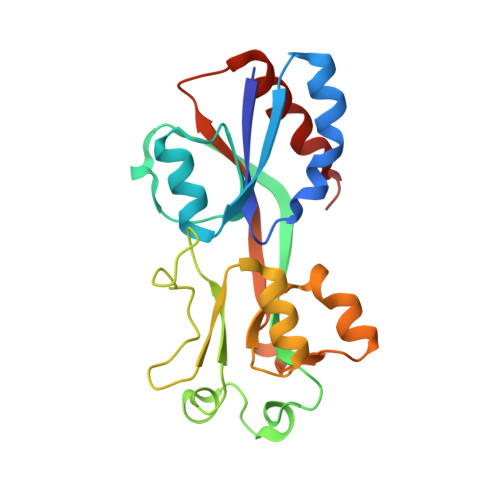
    Detection of landmines without harming personnel is a global issue. The bacterial transcription factor YhaJ selectively detects metabolites of explosives, and it can be used as a key component of DNT biosensors. However, the wild-type YhaJ has a binding affinity that is not sufficient for the detection of trace amounts of explosives leaked from landmines buried in the soil. Here, we report crystal structures of the effector-binding domain of YhaJ in both the apo- and effector-bound forms. A structural comparison of the two forms revealed that the loop above the primary effector-binding site significantly switches its conformation upon effector binding. The primary effector-binding site involves hydrophobic and polar interactions, having specificity to hydroxyl-substituted benzene compounds. The structures explain the mechanism of activity-enhancing mutations and provide information for the rational engineering of YhaJ biosensors for the sensitive detection of explosives.


  • Organizational Affiliation
    • Department of Bioengineering, College of Engineering, Hanyang University, Seoul 04673, Republic of Korea.

Macromolecule Content 

  • Total Structure Weight: 370.32 kDa 
  • Atom Count: 25,530 
  • Modeled Residue Count: 3,245 
  • Deposited Residue Count: 3,312 
  • Unique protein chains: 1

Macromolecules

Find similar proteins by:|  3D Structure
Entity ID: 1
MoleculeChains  Sequence LengthOrganismDetailsImage
HTH-type transcriptional regulator YhaJ
A, B, C, D, E
A, B, C, D, E, F, G, H, I, J, K, L, M, N, O, P
207Escherichia coliMutation(s): 0 
Gene Names: yhaJ
UniProt
Find proteins for P67660 (Escherichia coli (strain K12))
Explore P67660 
Go to UniProtKB:  P67660
Entity Groups
Sequence Clusters30% Identity50% Identity70% Identity90% Identity95% Identity100% Identity
UniProt GroupP67660
Sequence Annotations
Expand
Reference Sequence

Small Molecules

Ligands 1 Unique
IDChains Name / Formula / InChI Key2D Diagram3D Interactions
NA

Query on NA


Download:Ideal Coordinates CCD File
AA [auth L]
BA [auth M]
CA [auth N]
DA [auth O]
EA [auth P]
AA [auth L],
BA [auth M],
CA [auth N],
DA [auth O],
EA [auth P],
Q [auth A],
R [auth A],
S [auth C],
T [auth D],
U [auth F],
V [auth G],
W [auth H],
X [auth I],
Y [auth J],
Z [auth L]
SODIUM ION
Na
FKNQFGJONOIPTF-UHFFFAOYSA-N

Experimental Data & Validation

Experimental Data

  • Method: X-RAY DIFFRACTION
  • Resolution: 2.64 Å
  • R-Value Free:  0.236 (Depositor), 0.203 (DCC) 
  • R-Value Work:  0.177 (Depositor), 0.157 (DCC) 
Space Group: H 3
Unit Cell:
Length ( Å )Angle ( ˚ )
a = 215.193α = 90
b = 215.193β = 90
c = 263.409γ = 120
Software Package:
Software NamePurpose
REFMACrefinement
HKL-2000data reduction
HKL-2000data scaling
PHASERphasing

Structure Validation

View Full Validation Report


Entry History 

& Funding Information

Deposition Data


Funding OrganizationLocationGrant Number
National Research Foundation (NRF, Korea)Korea, Republic Of--

Revision History  (Full details and data files)

  • Version 1.0: 2023-10-25
    Type: Initial release
  • Version 1.1: 2024-03-06
    Changes: Source and taxonomy
  • Version 1.2: 2026-03-04
    Changes: Refinement description, Structure summary