8GUW

Structure of Aurora Kinase A in complex with activator peptide


Experimental Data Snapshot

  • Method: X-RAY DIFFRACTION
  • Resolution: 2.70 Å
  • R-Value Free: 0.258 
  • R-Value Work: 0.212 
  • R-Value Observed: 0.216 

wwPDB Validation   3D Report Full Report


Ligand Structure Quality Assessment 


This is version 1.1 of the entry. See complete history


Literature

Structural basis for CEP192-mediated regulation of centrosomal AURKA.

Park, J.G.Jeon, H.Shin, S.Song, C.Lee, H.Kim, N.K.Kim, E.E.Hwang, K.Y.Lee, B.J.Lee, I.G.

(2023) Sci Adv 9: eadf8582-eadf8582

  • DOI: https://doi.org/10.1126/sciadv.adf8582
  • Primary Citation of Related Structures:  
    8GUW

  • PubMed Abstract: 

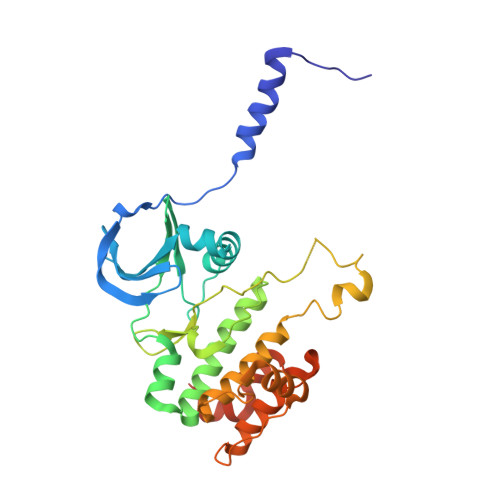
    Aurora kinase A (AURKA) performs critical functions in mitosis. Thus, the activity and subcellular localization of AURKA are tightly regulated and depend on diverse factors including interactions with the multiple binding cofactors. How these different cofactors regulate AURKA to elicit different levels of activity at distinct subcellular locations and times is poorly understood. Here, we identified a conserved region of CEP192, the major cofactor of AURKA, that mediates the interaction with AURKA. Quantitative binding studies were performed to map the interactions of a conserved helix (Helix-1) within CEP192. The crystal structure of Helix-1 bound to AURKA revealed a distinct binding site that is different from other cofactor proteins such as TPX2. Inhibiting the interaction between Helix-1 and AURKA in cells led to the mitotic defects, demonstrating the importance of the interaction. Collectively, we revealed a structural basis for the CEP192-mediated AURKA regulation at the centrosome, which is distinct from TPX2-mediated regulation on the spindle microtubule.


  • Organizational Affiliation

    Biomedical Research Division, Korea Institute of Science and Technology, Seoul 02792, South Korea.


Macromolecules
Find similar proteins by:  (by identity cutoff)  |  3D Structure
Entity ID: 1
MoleculeChains Sequence LengthOrganismDetailsImage
Peptide from Centrosomal protein of 192 kDa,Aurora kinase A
A, B, C
304Homo sapiensMutation(s): 2 
Gene Names: 
EC: 2.7.11.1
UniProt & NIH Common Fund Data Resources
Find proteins for Q8TEP8 (Homo sapiens)
Explore Q8TEP8 
Go to UniProtKB:  Q8TEP8
PHAROS:  Q8TEP8
GTEx:  ENSG00000101639 
Find proteins for O14965 (Homo sapiens)
Explore O14965 
Go to UniProtKB:  O14965
PHAROS:  O14965
GTEx:  ENSG00000087586 
Entity Groups  
Sequence Clusters30% Identity50% Identity70% Identity90% Identity95% Identity100% Identity
UniProt GroupsO14965Q8TEP8
Sequence Annotations
Expand
  • Reference Sequence
Experimental Data & Validation

Experimental Data

  • Method: X-RAY DIFFRACTION
  • Resolution: 2.70 Å
  • R-Value Free: 0.258 
  • R-Value Work: 0.212 
  • R-Value Observed: 0.216 
  • Space Group: P 21 21 21
Unit Cell:
Length ( Å )Angle ( ˚ )
a = 64.859α = 90
b = 78.695β = 90
c = 185.634γ = 90
Software Package:
Software NamePurpose
PHENIXrefinement
PDB_EXTRACTdata extraction
HKL-2000data reduction
HKL-2000data scaling
PHENIXphasing

Structure Validation

View Full Validation Report


Ligand Structure Quality Assessment 


Entry History & Funding Information

Deposition Data


Funding OrganizationLocationGrant Number
National Research Foundation (NRF, Korea)Korea, Republic Of--

Revision History  (Full details and data files)

  • Version 1.0: 2023-05-31
    Type: Initial release
  • Version 1.1: 2023-11-29
    Changes: Data collection, Refinement description