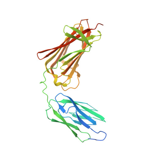
8GSX

X-ray structure of Clostridium perfringens pili protein B collagen-binding domains


Experimental Data Snapshot

  • Method: X-RAY DIFFRACTION
  • Resolution: 2.86 Å
  • R-Value Free: 0.286 
  • R-Value Work: 0.252 
  • R-Value Observed: 0.254 

wwPDB Validation   3D Report Full Report


Ligand Structure Quality Assessment 


This is version 1.0 of the entry. See complete history


Literature

Structural and biochemical characterization of Clostridium perfringens pili protein B collagen-binding domains.

Tamai, E.Yamada, M.Ishida, T.Arimura, N.Matsunami, R.Sekiya, H.Kamitori, S.

(2023) FEBS Lett 597: 1345-1354

  • DOI: https://doi.org/10.1002/1873-3468.14626
  • Primary Citation of Related Structures:  
    8GSX, 8GSY

  • PubMed Abstract: 

    Sortase-mediated pili are flexible rod proteins composed of major and minor/tip pilins, playing important roles in the initial adhesion of bacterial cells to host tissues. The pilus shaft is formed by covalent polymerization of major pilins, and the minor/tip pilin is covalently attached to the tip of the shaft involved in adhesion to the host cell. The Gram-positive bacterium Clostridium perfringens has a major pilin, and a minor/tip pilin (CppB) with the collagen-binding motif. Here, we report X-ray structures of CppB collagen-binding domains, collagen-binding assays and mutagenesis analysis, demonstrating that CppB collagen-binding domains adopt an L-shaped structure in open form, and that a small β-sheet unique to CppB provides a scaffold for a favourable binding site for collagen peptide.


  • Organizational Affiliation

    Department of Infectious Disease, College of Pharmaceutical Sciences, Matsuyama University, Japan.


Macromolecules
Find similar proteins by:  (by identity cutoff)  |  3D Structure
Entity ID: 1
MoleculeChains Sequence LengthOrganismDetailsImage
pili protein335Clostridium perfringens str. 13Mutation(s): 0 
Gene Names: CPE0155
UniProt
Find proteins for Q8XP11 (Clostridium perfringens (strain 13 / Type A))
Explore Q8XP11 
Go to UniProtKB:  Q8XP11
Entity Groups  
Sequence Clusters30% Identity50% Identity70% Identity90% Identity95% Identity100% Identity
UniProt GroupQ8XP11
Sequence Annotations
Expand
  • Reference Sequence
Experimental Data & Validation

Experimental Data

  • Method: X-RAY DIFFRACTION
  • Resolution: 2.86 Å
  • R-Value Free: 0.286 
  • R-Value Work: 0.252 
  • R-Value Observed: 0.254 
  • Space Group: P 65 2 2
Unit Cell:
Length ( Å )Angle ( ˚ )
a = 127.39α = 90
b = 127.39β = 90
c = 163.16γ = 120
Software Package:
Software NamePurpose
REFMACrefinement
XDSdata reduction
XDSdata scaling
PHENIXphasing

Structure Validation

View Full Validation Report


Ligand Structure Quality Assessment 


Entry History & Funding Information

Deposition Data


Funding OrganizationLocationGrant Number
Japan Society for the Promotion of Science (JSPS)Japan18K0687

Revision History  (Full details and data files)

  • Version 1.0: 2023-06-07
    Type: Initial release