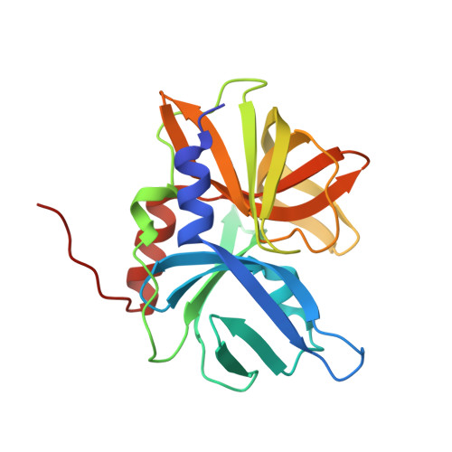
Allosteric regulation of Senecavirus A 3Cpro proteolytic activity by an endogenous phospholipid.
Zhao, H.F., Meng, L., Geng, Z., Gao, Z.Q., Dong, Y.H., Wang, H.W., Zhang, H.(2023) PLoS Pathog 19: e1011411-e1011411
- PubMed: 37253057 Search on PubMedSearch on PubMed Central
- DOI: https://doi.org/10.1371/journal.ppat.1011411
- Primary Citation Related Structures:
8GOT, 8GPH - PubMed Abstract:
Seneca virus A (SVA) is an emerging novel picornavirus that has recently been identified as the causative agent of many cases of porcine vesicular diseases in multiple countries. In addition to cleavage of viral polyprotein, the viral 3C protease (3Cpro) plays an important role in the regulation of several physiological processes involved in cellular antiviral responses by cleaving critical cellular proteins. Through a combination of crystallography, untargeted lipidomics, and immunoblotting, we identified the association of SVA 3Cpro with an endogenous phospholipid molecule, which binds to a unique region neighboring the proteolytic site of SVA 3Cpro. Our lipid-binding assays showed that SVA 3Cpro displayed preferred binding to cardiolipin (CL), followed by phosphoinositol-4-phosphate (PI4P) and sulfatide. Importantly, we found that the proteolytic activity of SVA 3Cpro was activated in the presence of the phospholipid, and the enzymatic activity is inhibited when the phospholipid-binding capacity decreased. Interestingly, in the wild-type SVA 3Cpro-substrate peptide structure, the cleavage residue cannot form a covalent binding to the catalytic cysteine residue to form the acyl-enzyme intermediate observed in several picornaviral 3Cpro structures. We observed a decrease in infectivity titers of SVA mutants harboring mutations that impaired the lipid-binding ability of 3Cpro, indicating a positive regulation of SVA infection capacity mediated by phospholipids. Our findings reveal a mutual regulation between the proteolytic activity and phospholipid-binding capacity in SVA 3Cpro, suggesting that endogenous phospholipid may function as an allosteric activator that regulate the enzyme's proteolytic activity during infection.
- School of Life Sciences, University of Science and Technology of China, Hefei, China.
Organizational Affiliation: