8GNI | pdb_00008gni

Human SARM1 bounded with NMN and Nanobody-C6, Conformation 1


Experimental Data Snapshot

  • Method: ELECTRON MICROSCOPY
  • Resolution: 3.74 Å
  • Aggregation State: PARTICLE 
  • Reconstruction Method: SINGLE PARTICLE 

Starting Models: experimental
View more details

wwPDB Validation 3D Report Full Report

Validation slider image for 8GNI

This is version 1.1 of the entry. See complete history

Literature

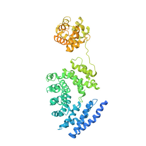
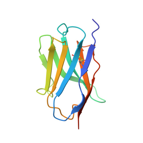
A conformation-specific nanobody targeting the nicotinamide mononucleotide-activated state of SARM1.

Hou, Y.N.Cai, Y.Li, W.H.He, W.M.Zhao, Z.Y.Zhu, W.J.Wang, Q.Mai, X.Liu, J.Lee, H.C.Stjepanovic, G.Zhang, H.Zhao, Y.J.

(2022) Nat Commun 13: 7898-7898

  • DOI: https://doi.org/10.1038/s41467-022-35581-y
  • Primary Citation Related Structures: 
    8GNI, 8GNJ, 8GQ5

  • PubMed Abstract: 

    Sterile alpha (SAM) and Toll/interleukin-1 receptor (TIR) motif containing 1 (SARM1) is an autoinhibitory NAD-consuming enzyme that is activated by the accumulation of nicotinamide mononucleotide (NMN) during axonal injury. Its activation mechanism is not fully understood. Here, we generate a nanobody, Nb-C6, that specifically recognizes NMN-activated SARM1. Nb-C6 stains only the activated SARM1 in cells stimulated with CZ-48, a permeant mimetic of NMN, and partially activates SARM1 in vitro and in cells. Cryo-EM of NMN/SARM1/Nb-C6 complex shows an octameric structure with ARM domains bending significantly inward and swinging out together with TIR domains. Nb-C6 binds to SAM domain of the activated SARM1 and stabilized its ARM domain. Mass spectrometry analyses indicate that the activated SARM1 in solution is highly dynamic and that the neighboring TIRs form transient dimers via the surface close to one BB loop. We show that Nb-C6 is a valuable tool for studies of SARM1 activation.


  • Organizational Affiliation
    • State Key Laboratory of Chemical Oncogenomics, Key Laboratory of Chemical Genomics, Peking University Shenzhen Graduate School, Shenzhen, 518055, China.

Macromolecule Content 

  • Total Structure Weight: 172.48 kDa 
  • Atom Count: 5,832 
  • Modeled Residue Count: 746 
  • Deposited Residue Count: 1,567 
  • Unique protein chains: 2

Macromolecules

Find similar proteins by:|  3D Structure
Entity ID: 1
MoleculeChains  Sequence LengthOrganismDetailsImage
NAD(+) hydrolase SARM1
A, B
724Homo sapiensMutation(s): 0 
Gene Names: SARM1KIAA0524SAMD2SARM
EC: 3.2.2.6 (PDB Primary Data), 3.2.2 (PDB Primary Data)
UniProt & NIH Common Fund Data Resources
Find proteins for Q6SZW1 (Homo sapiens)
Explore Q6SZW1 
Go to UniProtKB:  Q6SZW1
PHAROS:  Q6SZW1
GTEx:  ENSG00000004139 
Entity Groups
Sequence Clusters30% Identity50% Identity70% Identity90% Identity95% Identity100% Identity
UniProt GroupQ6SZW1
Sequence Annotations
Expand
Reference Sequence
Find similar proteins by:|  3D Structure
Entity ID: 2
MoleculeChains  Sequence LengthOrganismDetailsImage
Nanobody C6119Vicugna pacosMutation(s): 0 

Small Molecules

Ligands 1 Unique
IDChains Name / Formula / InChI Key2D Diagram3D Interactions
NMN
(Subject of Investigation/LOI)

Query on NMN


Download:Ideal Coordinates CCD File
D [auth A]BETA-NICOTINAMIDE RIBOSE MONOPHOSPHATE
C11 H16 N2 O8 P
DAYLJWODMCOQEW-TURQNECASA-O

Experimental Data & Validation

Experimental Data

  • Method: ELECTRON MICROSCOPY
  • Resolution: 3.74 Å
  • Aggregation State: PARTICLE 
  • Reconstruction Method: SINGLE PARTICLE 
EM Software:
TaskSoftware PackageVersion
MODEL REFINEMENTPHENIX1.16
RECONSTRUCTIONRELION3.1.2

Structure Validation

View Full Validation Report


Entry History 

& Funding Information

Deposition Data


Funding OrganizationLocationGrant Number
National Science Foundation (NSF, China)China--

Revision History  (Full details and data files)

  • Version 1.0: 2023-01-18
    Type: Initial release
  • Version 1.1: 2024-10-23
    Changes: Data collection, Refinement description, Structure summary