8GN6 | pdb_00008gn6

Crystallization of Sialidase from Porphyromonas gingivalis


Experimental Data Snapshot

  • Method: X-RAY DIFFRACTION
  • Resolution: 2.10 Å
  • R-Value Free: 
    0.216 (Depositor), 0.216 (DCC) 
  • R-Value Work: 
    0.173 (Depositor), 0.179 (DCC) 
  • R-Value Observed: 
    0.175 (Depositor) 

Starting Model: experimental
View more details

wwPDB Validation 3D Report Full Report

Validation slider image for 8GN6

This is version 1.1 of the entry. See complete history

Literature

Structural and enzymatic characterization of the sialidase SiaPG from Porphyromonas gingivalis.

Dong, W.B.Jiang, Y.L.Zhu, Z.L.Zhu, J.Li, Y.Xia, R.Zhou, K.

(2023) Acta Crystallogr F Struct Biol Commun 79: 87-94

  • DOI: https://doi.org/10.1107/S2053230X23001735
  • Primary Citation Related Structures: 
    8GN6

  • PubMed Abstract: 

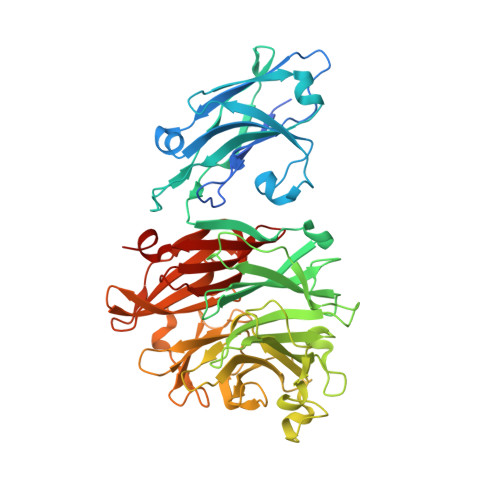
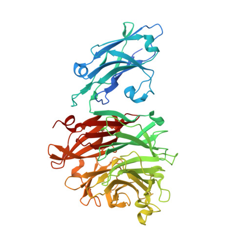
    The sialidases, which catalyze the hydrolysis of sialic acid from extracellular glycoconjugates, are a group of major virulence factors in various pathogenic bacteria. In Porphyromonas gingivalis, which causes human periodontal disease, sialidase contributes to bacterial pathogenesis via promoting the formation of biofilms and capsules, reducing the ability for macrophage clearance, and providing nutrients for bacterial colonization. Here, the crystal structure of the P. gingivalis sialidase SiaPG is reported at 2.1 Å resolution, revealing an N-terminal carbohydrate-binding domain followed by a canonical C-terminal catalytic domain. Simulation of the product sialic acid in the active-site pocket together with functional analysis enables clear identification of the key residues that are required for substrate binding and catalysis. Moreover, structural comparison with other sialidases reveals distinct features of the active-site pocket which might confer substrate specificity. These findings provide the structural basis for the further design and optimization of effective inhibitors to target SiaPG to fight against P. gingivalis-derived oral diseases.


  • Organizational Affiliation
    • Department of Stomatology, The Second Affiliated Hospital of Anhui Medical University, Hefei, Anhui 230601, People's Republic of China.

Macromolecule Content 

  • Total Structure Weight: 175.38 kDa 
  • Atom Count: 12,483 
  • Modeled Residue Count: 1,483 
  • Deposited Residue Count: 1,576 
  • Unique protein chains: 2

Macromolecules

Find similar proteins by:|  3D Structure
Entity ID: 1
MoleculeChains  Sequence LengthOrganismDetailsImage
Sialidase
A, B
526Porphyromonas gingivalisMutation(s): 0 
Gene Names: PGIN_13-1_00174
Find similar proteins by:|  3D Structure
Entity ID: 2
MoleculeChains  Sequence LengthOrganismDetailsImage
Sialidase524Porphyromonas gingivalisMutation(s): 0 
Gene Names: PGIN_13-1_00174

Small Molecules

Experimental Data & Validation

Experimental Data

  • Method: X-RAY DIFFRACTION
  • Resolution: 2.10 Å
  • R-Value Free:  0.216 (Depositor), 0.216 (DCC) 
  • R-Value Work:  0.173 (Depositor), 0.179 (DCC) 
  • R-Value Observed: 0.175 (Depositor) 
Space Group: P 31 2 1
Unit Cell:
Length ( Å )Angle ( ˚ )
a = 131.16α = 90
b = 131.16β = 90
c = 181.173γ = 120
Software Package:
Software NamePurpose
PHENIXrefinement
PDB_EXTRACTdata extraction
HKL-2000data reduction
HKL-2000data scaling
MLPHAREphasing

Structure Validation

View Full Validation Report


Entry History 

& Funding Information

Deposition Data

  • Released Date: 2023-04-19 
  • Deposition Author(s): Dong, W.B.

Funding OrganizationLocationGrant Number
Not funded--

Revision History  (Full details and data files)

  • Version 1.0: 2023-04-19
    Type: Initial release
  • Version 1.1: 2024-05-29
    Changes: Data collection