


Structure of methyltransferase
Zhang, H., Li, X.Z., Wen, Y.N.To be published.
Experimental Data Snapshot
Starting Model: in silico
View more details
wwPDB Validation 3D Report Full Report

Entity ID: 1 | |||||
|---|---|---|---|---|---|
| Molecule | Chains | Sequence Length | Organism | Details | Image |
| S-adenosylmethionine sensor upstream of mTORC1 | A [auth D] | 236 | Drosophila melanogaster | Mutation(s): 0 Gene Names: Samtor, CG3570 EC: 2.1.1 | 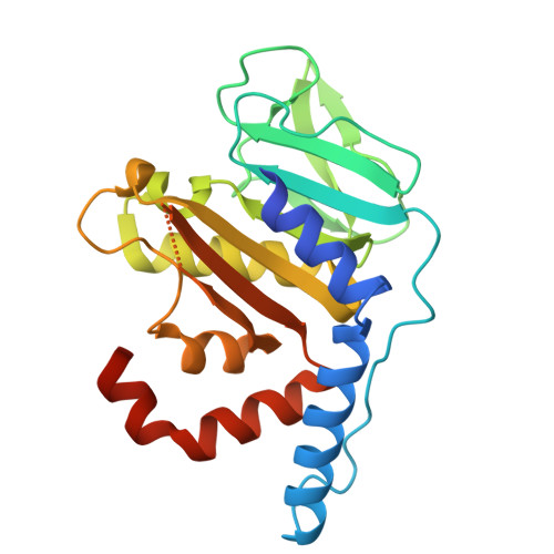 |
UniProt | |||||
Entity Groups | |||||
| Sequence Clusters | 30% Identity50% Identity70% Identity90% Identity95% Identity100% Identity | ||||
| UniProt Group | Q9W138 | ||||
Sequence AnnotationsExpand | |||||
Reference Sequence | |||||
| Length ( Å ) | Angle ( ˚ ) |
|---|---|
| a = 98.436 | α = 90 |
| b = 98.436 | β = 90 |
| c = 41.628 | γ = 120 |
| Software Name | Purpose |
|---|---|
| PHENIX | refinement |
| autoPROC | data reduction |
| autoPROC | data scaling |
| PHENIX | phasing |
| Funding Organization | Location | Grant Number |
|---|---|---|
| Other government | -- |


