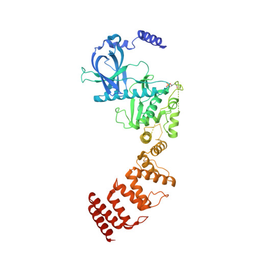
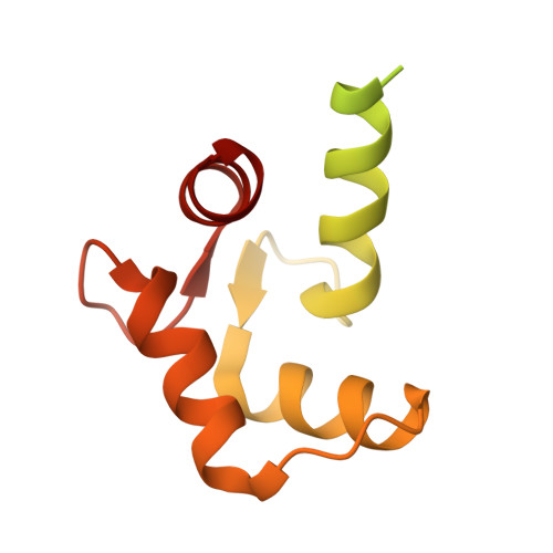
8GM4

Functional construct of the Eukaryotic elongation factor 2 kinase bound to an ATP-competitive inhibitor


Experimental Data Snapshot

  • Method: X-RAY DIFFRACTION
  • Resolution: 2.12 Å
  • R-Value Free: 0.238 
  • R-Value Work: 0.207 
  • R-Value Observed: 0.209 

wwPDB Validation   3D Report Full Report


Ligand Structure Quality Assessment 


This is version 1.2 of the entry. See complete history


Literature

Structure of the complex between calmodulin and a functional construct of eukaryotic elongation factor 2 kinase bound to an ATP-competitive inhibitor.

Piserchio, A.Isiorho, E.A.Dalby, K.N.Ghose, R.

(2023) J Biol Chem 299: 104813-104813

  • DOI: https://doi.org/10.1016/j.jbc.2023.104813
  • Primary Citation of Related Structures:  
    8GM4, 8GM5

  • PubMed Abstract: 

    The calmodulin-activated α-kinase, eukaryotic elongation factor 2 kinase (eEF-2K), serves as a master regulator of translational elongation by specifically phosphorylating and reducing the ribosome affinity of the guanosine triphosphatase, eukaryotic elongation factor 2 (eEF-2). Given its critical role in a fundamental cellular process, dysregulation of eEF-2K has been implicated in several human diseases, including those of the cardiovascular system, chronic neuropathies, and many cancers, making it a critical pharmacological target. In the absence of high-resolution structural information, high-throughput screening efforts have yielded small-molecule candidates that show promise as eEF-2K antagonists. Principal among these is the ATP-competitive pyrido-pyrimidinedione inhibitor, A-484954, which shows high specificity toward eEF-2K relative to a panel of "typical" protein kinases. A-484954 has been shown to have some degree of efficacy in animal models of several disease states. It has also been widely deployed as a reagent in eEF-2K-specific biochemical and cell-biological studies. However, given the absence of structural information, the precise mechanism of the A-484954-mediated inhibition of eEF-2K has remained obscure. Leveraging our identification of the calmodulin-activatable catalytic core of eEF-2K, and our recent determination of its long-elusive structure, here we present the structural basis for its specific inhibition by A-484954. This structure, which represents the first for an inhibitor-bound catalytic domain of a member of the α-kinase family, enables rationalization of the existing structure-activity relationship data for A-484954 variants and lays the groundwork for further optimization of this scaffold to attain enhanced specificity/potency against eEF-2K.


  • Organizational Affiliation

    Department of Chemistry and Biochemistry, The City College of New York, New York, New York, USA.


Macromolecules
Find similar proteins by:  (by identity cutoff)  |  3D Structure
Entity ID: 1
MoleculeChains Sequence LengthOrganismDetailsImage
Eukaryotic elongation factor 2 kinase531Homo sapiensMutation(s): 0 
Gene Names: EEF2K
EC: 2.7.11.20
UniProt & NIH Common Fund Data Resources
Find proteins for O00418 (Homo sapiens)
Explore O00418 
Go to UniProtKB:  O00418
PHAROS:  O00418
GTEx:  ENSG00000103319 
Entity Groups  
Sequence Clusters30% Identity50% Identity70% Identity90% Identity95% Identity100% Identity
UniProt GroupO00418
Sequence Annotations
Expand
  • Reference Sequence
Find similar proteins by:  (by identity cutoff)  |  3D Structure
Entity ID: 2
MoleculeChains Sequence LengthOrganismDetailsImage
Calmodulin-1148Homo sapiensMutation(s): 0 
Gene Names: CALM1CALMCAMCAM1
UniProt & NIH Common Fund Data Resources
Find proteins for P0DP23 (Homo sapiens)
Explore P0DP23 
Go to UniProtKB:  P0DP23
PHAROS:  P0DP23
GTEx:  ENSG00000198668 
Entity Groups  
Sequence Clusters30% Identity50% Identity70% Identity90% Identity95% Identity100% Identity
UniProt GroupP0DP23
Sequence Annotations
Expand
  • Reference Sequence
Small Molecules
Ligands 4 Unique
IDChains Name / Formula / InChI Key2D Diagram3D Interactions
ADP
Query on ADP

Download Ideal Coordinates CCD File 
C [auth A]ADENOSINE-5'-DIPHOSPHATE
C10 H15 N5 O10 P2
XTWYTFMLZFPYCI-KQYNXXCUSA-N
EKI (Subject of Investigation/LOI)
Query on EKI

Download Ideal Coordinates CCD File 
D [auth A]7-amino-1-cyclopropyl-3-ethyl-2,4-dioxo-1,2,3,4-tetrahydropyrido[2,3-d]pyrimidine-6-carboxamide
C13 H15 N5 O3
HWODCHXORCTEGU-UHFFFAOYSA-N
ZN
Query on ZN

Download Ideal Coordinates CCD File 
E [auth A]ZINC ION
Zn
PTFCDOFLOPIGGS-UHFFFAOYSA-N
CA
Query on CA

Download Ideal Coordinates CCD File 
F [auth B],
G [auth B]
CALCIUM ION
Ca
BHPQYMZQTOCNFJ-UHFFFAOYSA-N
Modified Residues  1 Unique
IDChains TypeFormula2D DiagramParent
TPO
Query on TPO
A
L-PEPTIDE LINKINGC4 H10 N O6 PTHR
Experimental Data & Validation

Experimental Data

  • Method: X-RAY DIFFRACTION
  • Resolution: 2.12 Å
  • R-Value Free: 0.238 
  • R-Value Work: 0.207 
  • R-Value Observed: 0.209 
  • Space Group: P 1 21 1
Unit Cell:
Length ( Å )Angle ( ˚ )
a = 80.544α = 90
b = 60.684β = 111.1
c = 88.704γ = 90
Software Package:
Software NamePurpose
PHENIXrefinement
autoPROCdata processing
Aimlessdata scaling
TRUNCATEdata processing
XDSdata reduction
PHASERphasing
Cootmodel building

Structure Validation

View Full Validation Report


Ligand Structure Quality Assessment 


Entry History & Funding Information

Deposition Data


Funding OrganizationLocationGrant Number
National Institutes of Health/National Institute of General Medical Sciences (NIH/NIGMS)United StatesGM123252

Revision History  (Full details and data files)

  • Version 1.0: 2023-05-31
    Type: Initial release
  • Version 1.1: 2023-06-21
    Changes: Database references
  • Version 1.2: 2023-10-25
    Changes: Data collection