8GD2

Crystal structure of human cellular retinol binding protein 1 in complex with N-methyl-1-{3-[1-(4-methylphenyl)cyclopentyl]-1,2,4-oxadiazol-5-yl}-N-(2-thienylmethyl)methanamine

  • Classification: Retinol-binding protein
  • Organism(s): Homo sapiens
  • Expression System: Escherichia coli
  • Mutation(s): No 

  • Deposited: 2023-03-03 Released: 2023-10-04 
  • Deposition Author(s): Plau, J., Golczak, M.
  • Funding Organization(s): National Institutes of Health/National Eye Institute (NIH/NEI), National Institutes of Health/National Institute of Diabetes and Digestive and Kidney Disease (NIH/NIDDK)

Experimental Data Snapshot

  • Method: X-RAY DIFFRACTION
  • Resolution: 1.13 Å
  • R-Value Free: 0.183 
  • R-Value Work: 0.162 
  • R-Value Observed: 0.163 

wwPDB Validation   3D Report Full Report


Ligand Structure Quality Assessment 


This is version 1.1 of the entry. See complete history


Literature

Discovery of Nonretinoid Inhibitors of CRBP1: Structural and Dynamic Insights for Ligand-Binding Mechanisms.

Plau, J.Morgan, C.E.Fedorov, Y.Banerjee, S.Adams, D.J.Blaner, W.S.Yu, E.W.Golczak, M.

(2023) ACS Chem Biol 18: 2309-2323

  • DOI: https://doi.org/10.1021/acschembio.3c00402
  • Primary Citation of Related Structures:  
    8GD2, 8GDM, 8GEM, 8GEU, 8GEV, 8GEY

  • PubMed Abstract: 

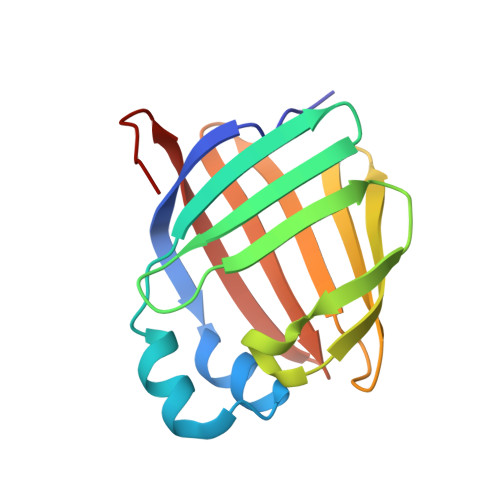
    The dysregulation of retinoid metabolism has been linked to prevalent ocular diseases including age-related macular degeneration and Stargardt disease. Modulating retinoid metabolism through pharmacological approaches holds promise for the treatment of these eye diseases. Cellular retinol-binding protein 1 (CRBP1) is the primary transporter of all- trans -retinol (atROL) in the eye, and its inhibition has recently been shown to protect mouse retinas from light-induced retinal damage. In this report, we employed high-throughput screening to identify new chemical scaffolds for competitive, nonretinoid inhibitors of CRBP1. To understand the mechanisms of interaction between CRBP1 and these inhibitors, we solved high-resolution X-ray crystal structures of the protein in complex with six selected compounds. By combining protein crystallography with hydrogen/deuterium exchange mass spectrometry, we quantified the conformational changes in CRBP1 caused by different inhibitors and correlated their magnitude with apparent binding affinities. Furthermore, using molecular dynamic simulations, we provided evidence for the functional significance of the "closed" conformation of CRBP1 in retaining ligands within the binding pocket. Collectively, our study outlines the molecular foundations for understanding the mechanism of high-affinity interactions between small molecules and CRBPs, offering a framework for the rational design of improved inhibitors for this class of lipid-binding proteins.


  • Organizational Affiliation

    Department of Chemistry, Thiel College, Greenville, Pennsylvania 16125, United States.


Macromolecules
Find similar proteins by:  (by identity cutoff)  |  3D Structure
Entity ID: 1
MoleculeChains Sequence LengthOrganismDetailsImage
Retinol-binding protein 1A [auth B]141Homo sapiensMutation(s): 0 
Gene Names: RBP1CRBP1
UniProt & NIH Common Fund Data Resources
Find proteins for P09455 (Homo sapiens)
Explore P09455 
Go to UniProtKB:  P09455
PHAROS:  P09455
GTEx:  ENSG00000114115 
Entity Groups  
Sequence Clusters30% Identity50% Identity70% Identity90% Identity95% Identity100% Identity
UniProt GroupP09455
Sequence Annotations
Expand
  • Reference Sequence
Small Molecules
Ligands 2 Unique
IDChains Name / Formula / InChI Key2D Diagram3D Interactions
Z5H (Subject of Investigation/LOI)
Query on Z5H

Download Ideal Coordinates CCD File 
B
N-methyl-1-{3-[1-(4-methylphenyl)cyclopentyl]-1,2,4-oxadiazol-5-yl}-N-[(thiophen-2-yl)methyl]methanamine
C21 H25 N3 O S
GPXYCFQNEZDPGQ-UHFFFAOYSA-N
BTB
Query on BTB

Download Ideal Coordinates CCD File 
C [auth B]2-[BIS-(2-HYDROXY-ETHYL)-AMINO]-2-HYDROXYMETHYL-PROPANE-1,3-DIOL
C8 H19 N O5
OWMVSZAMULFTJU-UHFFFAOYSA-N
Experimental Data & Validation

Experimental Data

  • Method: X-RAY DIFFRACTION
  • Resolution: 1.13 Å
  • R-Value Free: 0.183 
  • R-Value Work: 0.162 
  • R-Value Observed: 0.163 
  • Space Group: P 21 21 21
Unit Cell:
Length ( Å )Angle ( ˚ )
a = 37.329α = 90
b = 37.685β = 90
c = 101.544γ = 90
Software Package:
Software NamePurpose
PHENIXrefinement
XDSdata reduction
Aimlessdata scaling
PHASERphasing

Structure Validation

View Full Validation Report


Ligand Structure Quality Assessment 


Entry History & Funding Information

Deposition Data


Funding OrganizationLocationGrant Number
National Institutes of Health/National Eye Institute (NIH/NEI)United StatesEY023948
National Institutes of Health/National Institute of Diabetes and Digestive and Kidney Disease (NIH/NIDDK)United StatesDK122071

Revision History  (Full details and data files)

  • Version 1.0: 2023-10-04
    Type: Initial release
  • Version 1.1: 2023-11-01
    Changes: Database references