8GCY | pdb_00008gcy

Co-crystal structure of CBL-B in complex with N-Aryl isoindolin-1-one inhibitor


Experimental Data Snapshot

  • Method: X-RAY DIFFRACTION
  • Resolution: 1.81 Å
  • R-Value Free: 
    0.230 (Depositor), 0.238 (DCC) 
  • R-Value Work: 
    0.195 (Depositor), 0.202 (DCC) 
  • R-Value Observed: 
    0.197 (Depositor) 

Starting Model: experimental
View more details

wwPDB Validation 3D Report Full Report

Validation slider image for 8GCY

Ligand Structure Quality Assessment 


This is version 1.1 of the entry. See complete history

Literature

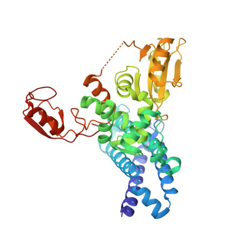
The co-crystal structure of Cbl-b and a small-molecule inhibitor reveals the mechanism of Cbl-b inhibition.

Kimani, S.W.Perveen, S.Szewezyk, M.Zeng, H.Dong, A.Li, F.Ghiabi, P.Li, Y.Chau, I.Arrowsmith, C.H.Barsyte-Lovejoy, D.Santhakumar, V.Vedadi, M.Halabelian, L.

(2023) Commun Biol 6: 1272-1272

  • DOI: https://doi.org/10.1038/s42003-023-05655-8
  • Primary Citation Related Structures: 
    8GCY

  • PubMed Abstract: 

    Cbl-b is a RING-type E3 ubiquitin ligase that is expressed in several immune cell lineages, where it negatively regulates the activity of immune cells. Cbl-b has specifically been identified as an attractive target for cancer immunotherapy due to its role in promoting an immunosuppressive tumor environment. A Cbl-b inhibitor, Nx-1607, is currently in phase I clinical trials for advanced solid tumor malignancies. Using a suite of biophysical and cellular assays, we confirm potent binding of C7683 (an analogue of Nx-1607) to the full-length Cbl-b and its N-terminal fragment containing the TKBD-LHR-RING domains. To further elucidate its mechanism of inhibition, we determined the co-crystal structure of Cbl-b with C7683, revealing the compound's interaction with both the TKBD and LHR, but not the RING domain. Here, we provide structural insights into a novel mechanism of Cbl-b inhibition by a small-molecule inhibitor that locks the protein in an inactive conformation by acting as an intramolecular glue.


  • Organizational Affiliation
    • Structural Genomics Consortium, University of Toronto, Toronto, ON, Canada.

Macromolecule Content 

  • Total Structure Weight: 45.98 kDa 
  • Atom Count: 3,233 
  • Modeled Residue Count: 376 
  • Deposited Residue Count: 391 
  • Unique protein chains: 1

Macromolecules

Find similar proteins by:|  3D Structure
Entity ID: 1
MoleculeChains  Sequence LengthOrganismDetailsImage
E3 ubiquitin-protein ligase CBL-B391Homo sapiensMutation(s): 0 
Gene Names: CBLBRNF56Nbla00127
EC: 2.3.2.27
UniProt & NIH Common Fund Data Resources
Find proteins for Q13191 (Homo sapiens)
Explore Q13191 
Go to UniProtKB:  Q13191
PHAROS:  Q13191
GTEx:  ENSG00000114423 
Entity Groups
Sequence Clusters30% Identity50% Identity70% Identity90% Identity95% Identity100% Identity
UniProt GroupQ13191
Sequence Annotations
Expand
Reference Sequence

Small Molecules

Ligands 5 Unique
IDChains Name / Formula / InChI Key2D Diagram3D Interactions
Z3N
(Subject of Investigation/LOI)

Query on Z3N


Download:Ideal Coordinates CCD File
D [auth A]2-{3-[(1s,3R)-3-methyl-1-(4-methyl-4H-1,2,4-triazol-3-yl)cyclobutyl]phenyl}-6-{[(3S)-3-methylpiperidin-1-yl]methyl}-4-(trifluoromethyl)-2,3-dihydro-1H-isoindol-1-one
C30 H34 F3 N5 O
HUOLMBXGHHSYHC-QWQFASRJSA-N
SO4

Query on SO4


Download:Ideal Coordinates CCD File
F [auth A]SULFATE ION
O4 S
QAOWNCQODCNURD-UHFFFAOYSA-L
ZN

Query on ZN


Download:Ideal Coordinates CCD File
B [auth A],
C [auth A]
ZINC ION
Zn
PTFCDOFLOPIGGS-UHFFFAOYSA-N
EDO

Query on EDO


Download:Ideal Coordinates CCD File
E [auth A]1,2-ETHANEDIOL
C2 H6 O2
LYCAIKOWRPUZTN-UHFFFAOYSA-N
UNX

Query on UNX


Download:Ideal Coordinates CCD File
G [auth A]UNKNOWN ATOM OR ION
X
Binding Affinity Annotations 
IDSourceBinding Affinity
Z3N BindingDB:  8GCY Kd: min: 3, max: 8 (nM) from 2 assay(s)
IC50: min: 5, max: 9 (nM) from 2 assay(s)

Experimental Data & Validation

Experimental Data

  • Method: X-RAY DIFFRACTION
  • Resolution: 1.81 Å
  • R-Value Free:  0.230 (Depositor), 0.238 (DCC) 
  • R-Value Work:  0.195 (Depositor), 0.202 (DCC) 
  • R-Value Observed: 0.197 (Depositor) 
Space Group: P 21 21 21
Unit Cell:
Length ( Å )Angle ( ˚ )
a = 47.937α = 90
b = 76.093β = 90
c = 105.464γ = 90
Software Package:
Software NamePurpose
HKL-3000data scaling
REFMACrefinement
HKL-3000data reduction
PHASERphasing

Structure Validation

View Full Validation Report


Ligand Structure Quality Assessment 


Entry History 

& Funding Information

Deposition Data


Funding OrganizationLocationGrant Number
Other private--

Revision History  (Full details and data files)

  • Version 1.0: 2023-03-22
    Type: Initial release
  • Version 1.1: 2024-04-03
    Changes: Data collection, Database references