8G89 | pdb_00008g89

HSD17B13 in complex with cofactor and inhibitor


Experimental Data Snapshot

  • Method: X-RAY DIFFRACTION
  • Resolution: 2.22 Å
  • R-Value Free: 
    0.228 (Depositor), 0.225 (DCC) 
  • R-Value Work: 
    0.209 (Depositor), 0.206 (DCC) 
  • R-Value Observed: 
    0.209 (Depositor) 

Starting Model: experimental
View more details

wwPDB Validation 3D Report Full Report

Validation slider image for 8G89

Ligand Structure Quality Assessment 


This is version 1.1 of the entry. See complete history

Literature

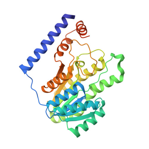
Structural basis of lipid-droplet localization of 17-beta-hydroxysteroid dehydrogenase 13.

Liu, S.Sommese, R.F.Nedoma, N.L.Stevens, L.M.Dutra, J.K.Zhang, L.Edmonds, D.J.Wang, Y.Garnsey, M.Clasquin, M.F.

(2023) Nat Commun 14: 5158-5158

  • DOI: https://doi.org/10.1038/s41467-023-40766-0
  • Primary Citation Related Structures: 
    8G84, 8G89, 8G93, 8G9V

  • PubMed Abstract: 

    Hydroxysteroid 17-beta-dehydrogenase 13 (HSD17B13) is a hepatic lipid droplet-associated enzyme that is upregulated in patients with non-alcoholic fatty liver disease. Recently, there have been several reports that predicted loss of function variants in HSD17B13 protect against the progression of steatosis to non-alcoholic steatohepatitis with fibrosis and hepatocellular carcinoma. Here we report crystal structures of full length HSD17B13 in complex with its NAD + cofactor, and with lipid/detergent molecules and small molecule inhibitors from two distinct series in the ligand binding pocket. These structures provide insights into a mechanism for lipid droplet-associated proteins anchoring to membranes as well as a basis for HSD17B13 variants disrupting function. Two series of inhibitors interact with the active site residues and the bound cofactor similarly, yet they occupy different paths leading to the active site. These structures provide ideas for structure-based design of inhibitors that may be used in the treatment of liver disease.


  • Organizational Affiliation
    • Medicine Design, Pfizer Inc, Groton, CT, 06340, USA. Shenping.Liu@pfizer.com.

Macromolecule Content 

  • Total Structure Weight: 72.45 kDa 
  • Atom Count: 4,750 
  • Modeled Residue Count: 579 
  • Deposited Residue Count: 630 
  • Unique protein chains: 1

Macromolecules

Find similar proteins by:|  3D Structure
Entity ID: 1
MoleculeChains  Sequence LengthOrganismDetailsImage
Hydroxysteroid 17-beta dehydrogenase 13
A, B
315Canis lupus familiarisMutation(s): 0 
Gene Names: HSD17B13
EC: 1.1.1.62
UniProt
Find proteins for A0A8I3RYU0 (Canis lupus familiaris)
Explore A0A8I3RYU0 
Go to UniProtKB:  A0A8I3RYU0
Entity Groups
Sequence Clusters30% Identity50% Identity70% Identity90% Identity95% Identity100% Identity
UniProt GroupA0A8I3RYU0
Sequence Annotations
Expand
Reference Sequence

Experimental Data & Validation

Experimental Data

  • Method: X-RAY DIFFRACTION
  • Resolution: 2.22 Å
  • R-Value Free:  0.228 (Depositor), 0.225 (DCC) 
  • R-Value Work:  0.209 (Depositor), 0.206 (DCC) 
  • R-Value Observed: 0.209 (Depositor) 
Space Group: P 21 21 2
Unit Cell:
Length ( Å )Angle ( ˚ )
a = 77.03α = 90
b = 186.46β = 90
c = 65.47γ = 90
Software Package:
Software NamePurpose
BUSTERrefinement
XDSdata reduction
autoPROCdata scaling
BUSTERphasing

Structure Validation

View Full Validation Report


Ligand Structure Quality Assessment 


Entry History 

& Funding Information

Deposition Data

  • Released Date: 2023-08-09 
  • Deposition Author(s): Liu, S.

Funding OrganizationLocationGrant Number
Not fundedUnited States--

Revision History  (Full details and data files)

  • Version 1.0: 2023-08-09
    Type: Initial release
  • Version 1.1: 2023-09-06
    Changes: Data collection, Database references