8G33 | pdb_00008g33

Activated form of a CDCL long protein


Experimental Data Snapshot

  • Method: X-RAY DIFFRACTION
  • Resolution: 2.49 Å
  • R-Value Free: 
    0.248 (Depositor), 0.250 (DCC) 
  • R-Value Work: 
    0.207 (Depositor), 0.210 (DCC) 
  • R-Value Observed: 
    0.210 (Depositor) 

Starting Model: experimental
View more details

wwPDB Validation   3D Report Full Report


This is version 1.1 of the entry. See complete history


Literature

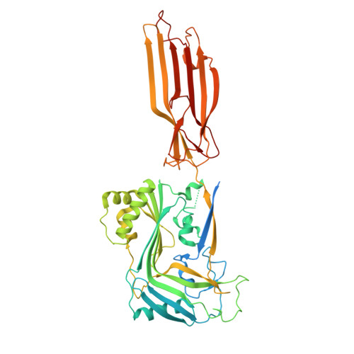
Structural basis for the pore-forming activity of a complement-like toxin.

Johnstone, B.A.Christie, M.P.Joseph, R.Morton, C.J.Brown, H.G.Hanssen, E.Sanford, T.C.Abrahamsen, H.L.Tweten, R.K.Parker, M.W.

(2025) Sci Adv 11: eadt2127-eadt2127

  • DOI: https://doi.org/10.1126/sciadv.adt2127
  • Primary Citation of Related Structures:  
    8G33, 9CCP, 9CCQ

  • PubMed Abstract: 

    Pore-forming proteins comprise a highly diverse group of proteins exemplified by the membrane attack complex/perforin (MACPF), cholesterol-dependent cytolysin (CDC), and gasdermin superfamilies, which all form gigantic pores (>150 angstroms). A recently found family of pore-forming toxins, called CDC-like proteins (CDCLs), are wide-spread in gut microbes and are a prevalent means of antibacterial antagonism. However, the structural aspects of how CDCLs assemble a pore remain a mystery. Here, we report the crystal structure of a proteolytically activated CDCL and cryo-electron microscopy structures of a prepore-like intermediate and a transmembrane pore providing detailed snapshots across the entire pore-forming pathway. These studies reveal a sophisticated array of regulatory features to ensure productive pore formation, and, thus, CDCLs straddle the MACPF, CDC, and gasdermin lineages of the giant pore superfamilies.


  • Organizational Affiliation
    • Department of Biochemistry and Pharmacology, Bio21 Molecular Science and Biotechnology Institute, University of Melbourne, Parkville, VIC 3010, Australia.

Macromolecules
Find similar proteins by:  (by identity cutoff)  |  3D Structure
Entity ID: 1
MoleculeChains Sequence LengthOrganismDetailsImage
Hemolysin497Elizabethkingia anophelis Ag1Mutation(s): 0 
Gene Names: ply_2AYC66_13765BAY09_14710NCTC10586_03727
UniProt
Find proteins for A0A1T3IZT7 (Elizabethkingia anophelis)
Explore A0A1T3IZT7 
Go to UniProtKB:  A0A1T3IZT7
Entity Groups  
Sequence Clusters30% Identity50% Identity70% Identity90% Identity95% Identity100% Identity
UniProt GroupA0A1T3IZT7
Sequence Annotations
Expand
  • Reference Sequence
Experimental Data & Validation

Experimental Data

  • Method: X-RAY DIFFRACTION
  • Resolution: 2.49 Å
  • R-Value Free:  0.248 (Depositor), 0.250 (DCC) 
  • R-Value Work:  0.207 (Depositor), 0.210 (DCC) 
  • R-Value Observed: 0.210 (Depositor) 
Space Group: I 1 2 1
Unit Cell:
Length ( Å )Angle ( ˚ )
a = 61.725α = 90
b = 24.451β = 93.19
c = 300.191γ = 90
Software Package:
Software NamePurpose
XDSdata reduction
Aimlessdata scaling
PHASERphasing
PHENIXrefinement
Cootmodel building

Structure Validation

View Full Validation Report


Entry History & Funding Information

Deposition Data


Funding OrganizationLocationGrant Number
Australian Research Council (ARC)AustraliaDP200102871
Australian Research Council (ARC)AustraliaDP160101874

Revision History  (Full details and data files)

  • Version 1.0: 2024-02-07
    Type: Initial release
  • Version 1.1: 2025-04-09
    Changes: Database references, Structure summary