8G2Y

Cryo-EM structure of ADGRF1 coupled to miniGs/q


Experimental Data Snapshot

  • Method: ELECTRON MICROSCOPY
  • Resolution: 3.44 Å
  • Aggregation State: PARTICLE 
  • Reconstruction Method: SINGLE PARTICLE 

wwPDB Validation   3D Report Full Report


This is version 1.0 of the entry. See complete history


Literature

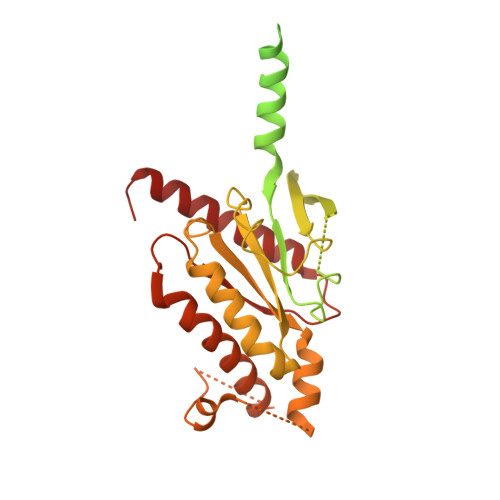
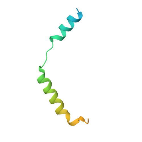
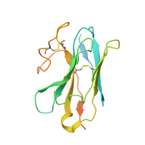
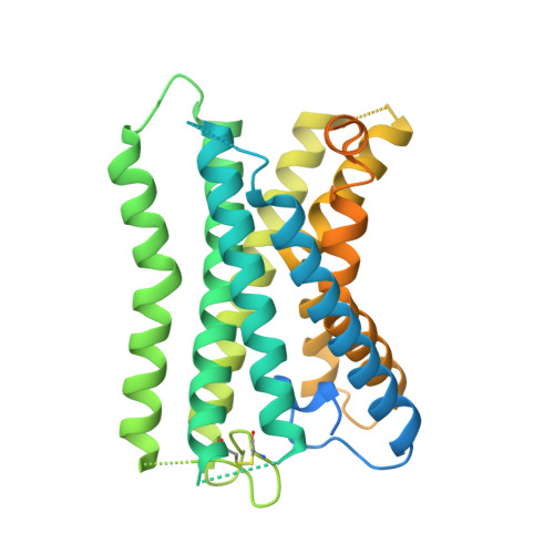
Tethered agonist activated ADGRF1 structure and signalling analysis reveal basis for G protein coupling.

Jones, D.T.D.Dates, A.N.Rawson, S.D.Burruss, M.M.Lipper, C.H.Blacklow, S.C.

(2023) Nat Commun 14: 2490-2490

  • DOI: https://doi.org/10.1038/s41467-023-38083-7
  • Primary Citation of Related Structures:  
    8G2Y

  • PubMed Abstract: 

    Adhesion G Protein Coupled Receptors (aGPCRs) have evolved an activation mechanism to translate extracellular force into liberation of a tethered agonist (TA) to effect cell signalling. We report here that ADGRF1 can signal through all major G protein classes and identify the structural basis for a previously reported Gα q preference by cryo-EM. Our structure shows that Gα q preference in ADGRF1 may derive from tighter packing at the conserved F569 of the TA, altering contacts between TM helix I and VII, with a concurrent rearrangement of TM helix VII and helix VIII at the site of Gα recruitment. Mutational studies of the interface and of contact residues within the 7TM domain identify residues critical for signalling, and suggest that Gα s signalling is more sensitive to mutation of TA or binding site residues than Gα q . Our work advances the detailed molecular understanding of aGPCR TA activation, identifying features that potentially explain preferential signal modulation.


  • Organizational Affiliation

    Department of Biological Chemistry and Molecular Pharmacology, Blavatnik Institute, Harvard Medical School, Boston, MA, 02115, USA. daniel_jones@hms.harvard.edu.


Macromolecules
Find similar proteins by:  (by identity cutoff)  |  3D Structure
Entity ID: 1
MoleculeChains Sequence LengthOrganismDetailsImage
MiniG alpha s/q chimera423Homo sapiensMutation(s): 0 
Entity Groups  
Sequence Clusters30% Identity50% Identity70% Identity90% Identity95% Identity100% Identity
Sequence Annotations
Expand
  • Reference Sequence
Find similar proteins by:  (by identity cutoff)  |  3D Structure
Entity ID: 2
MoleculeChains Sequence LengthOrganismDetailsImage
Guanine nucleotide-binding protein G(I)/G(S)/G(T) subunit beta-1358Homo sapiensMutation(s): 0 
Gene Names: GNB1
UniProt & NIH Common Fund Data Resources
Find proteins for P62873 (Homo sapiens)
Explore P62873 
Go to UniProtKB:  P62873
PHAROS:  P62873
GTEx:  ENSG00000078369 
Entity Groups  
Sequence Clusters30% Identity50% Identity70% Identity90% Identity95% Identity100% Identity
UniProt GroupP62873
Sequence Annotations
Expand
  • Reference Sequence
Find similar proteins by:  (by identity cutoff)  |  3D Structure
Entity ID: 3
MoleculeChains Sequence LengthOrganismDetailsImage
Guanine nucleotide-binding protein G(I)/G(S)/G(O) subunit gamma-2C [auth G]71Homo sapiensMutation(s): 0 
Gene Names: GNG2
Membrane Entity: Yes 
UniProt & NIH Common Fund Data Resources
Find proteins for P59768 (Homo sapiens)
Explore P59768 
Go to UniProtKB:  P59768
PHAROS:  P59768
GTEx:  ENSG00000186469 
Entity Groups  
Sequence Clusters30% Identity50% Identity70% Identity90% Identity95% Identity100% Identity
UniProt GroupP59768
Sequence Annotations
Expand
  • Reference Sequence
Find similar proteins by:  (by identity cutoff)  |  3D Structure
Entity ID: 4
MoleculeChains Sequence LengthOrganismDetailsImage
Nanobody 35D [auth N]181Lama glamaMutation(s): 0 
Entity Groups  
Sequence Clusters30% Identity50% Identity70% Identity90% Identity95% Identity100% Identity
Sequence Annotations
Expand
  • Reference Sequence
Find similar proteins by:  (by identity cutoff)  |  3D Structure
Entity ID: 5
MoleculeChains Sequence LengthOrganismDetailsImage
Adhesion G-protein-coupled receptor F1E [auth R]411Homo sapiensMutation(s): 0 
Gene Names: ADGRF1
Membrane Entity: Yes 
UniProt & NIH Common Fund Data Resources
Find proteins for Q5T601 (Homo sapiens)
Explore Q5T601 
Go to UniProtKB:  Q5T601
PHAROS:  Q5T601
GTEx:  ENSG00000153292 
Entity Groups  
Sequence Clusters30% Identity50% Identity70% Identity90% Identity95% Identity100% Identity
UniProt GroupQ5T601
Sequence Annotations
Expand
  • Reference Sequence
Small Molecules
Ligands 1 Unique
IDChains Name / Formula / InChI Key2D Diagram3D Interactions
LPC (Subject of Investigation/LOI)
Query on LPC

Download Ideal Coordinates CCD File 
F [auth R][1-MYRISTOYL-GLYCEROL-3-YL]PHOSPHONYLCHOLINE
C22 H47 N O7 P
VXUOFDJKYGDUJI-OAQYLSRUSA-O
Experimental Data & Validation

Experimental Data

  • Method: ELECTRON MICROSCOPY
  • Resolution: 3.44 Å
  • Aggregation State: PARTICLE 
  • Reconstruction Method: SINGLE PARTICLE 

Structure Validation

View Full Validation Report


Entry History & Funding Information

Deposition Data


Funding OrganizationLocationGrant Number
National Institutes of Health/National Cancer Institute (NIH/NCI)United States1R35 CA220340

Revision History  (Full details and data files)

  • Version 1.0: 2023-05-10
    Type: Initial release