Development of Novel Small-Molecule Activators of Pyruvate Kinase Muscle Isozyme 2, PKM2, to Reduce Photoreceptor Apoptosis.
Wubben, T.J., Chaudhury, S., Watch, B.T., Stuckey, J.A., Weh, E., Fernando, R., Goswami, M., Pawar, M., Rech, J.C., Besirli, C.G.(2023) Pharmaceuticals (Basel) 16
- PubMed: 37242488 Search on PubMedSearch on PubMed Central
- DOI: https://doi.org/10.3390/ph16050705
- Primary Citation Related Structures:
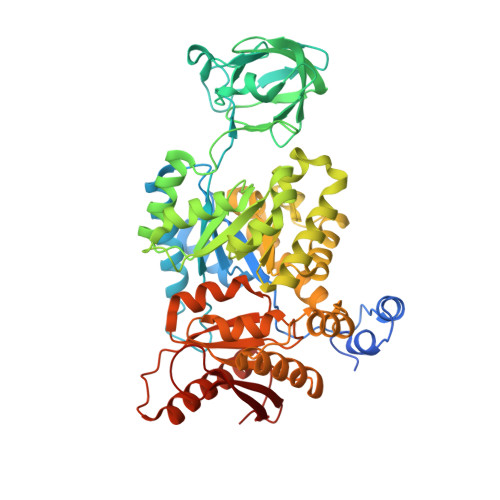
8G2E - PubMed Abstract:
Treatment options are lacking to prevent photoreceptor death and subsequent vision loss. Previously, we demonstrated that reprogramming metabolism via the pharmacologic activation of PKM2 is a novel photoreceptor neuroprotective strategy. However, the features of the tool compound used in those studies, ML-265, preclude its advancement as an intraocular, clinical candidate. This study sought to develop the next generation of small-molecule PKM2 activators, aimed specifically for delivery into the eye. Compounds were developed that replaced the thienopyrrolopyridazinone core of ML-265 and modified the aniline and methyl sulfoxide functional groups. Compound 2 demonstrated that structural changes to the ML-265 scaffold are tolerated from a potency and efficacy standpoint, allow for a similar binding mode to the target, and circumvent apoptosis in models of outer retinal stress. To overcome the low solubility and problematic functional groups of ML-265, compound 2 's efficacious and versatile core structure for the incorporation of diverse functional groups was then utilized to develop novel PKM2 activators with improved solubility, lack of structural alerts, and retained potency. No other molecules are in the pharmaceutical pipeline for the metabolic reprogramming of photoreceptors. Thus, this study is the first to cultivate the next generation of novel, structurally diverse, small-molecule PKM2 activators for delivery into the eye.
- Department of Ophthalmology and Visual Sciences, Kellogg Eye Center, University of Michigan, Ann Arbor, MI 48105, USA.
Organizational Affiliation: