8G25

Crystal Structure of Cathepsin-G and Neutrophil Elastase Inhibited by S. aureus EapH2 at pH 7.5


Experimental Data Snapshot

  • Method: X-RAY DIFFRACTION
  • Resolution: 1.80 Å
  • R-Value Free: 0.283 
  • R-Value Work: 0.248 
  • R-Value Observed: 0.249 

wwPDB Validation   3D Report Full Report


This is version 1.3 of the entry. See complete history


Literature

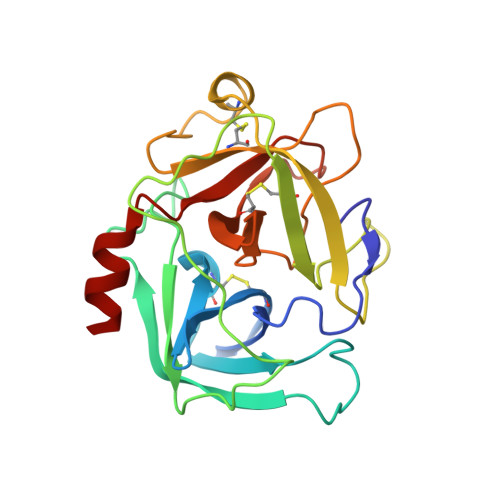
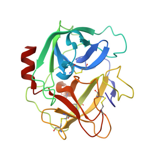
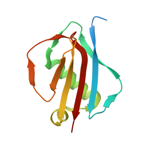
Simultaneous inhibition of two neutrophil serine proteases by the S. aureus innate immune evasion protein EapH2.

Mishra, N.Herdendorf, T.J.Prakash, O.Geisbrecht, B.V.

(2023) J Biol Chem 299: 104878-104878

  • DOI: https://doi.org/10.1016/j.jbc.2023.104878
  • Primary Citation of Related Structures:  
    8G24, 8G25, 8G26, 8GDG, 8GDH

  • PubMed Abstract: 

    Extracellular adherence protein domain (EAP) proteins are high-affinity, selective inhibitors of neutrophil serine proteases (NSP), including cathepsin-G (CG) and neutrophil elastase (NE). Most Staphylococcus aureus isolates encode for two EAPs, EapH1 and EapH2, that contain a single functional domain and share 43% identity with one another. Although structure/function investigations from our group have shown that EapH1 uses a globally similar binding mode to inhibit CG and NE, NSP inhibition by EapH2 is incompletely understood due to a lack of NSP/EapH2 cocrystal structures. To address this limitation, we further studied NSP inhibition by EapH2 in comparison with EapH1. Like its effects on NE, we found that EapH2 is a reversible, time-dependent, and low nanomolar affinity inhibitor of CG. We characterized an EapH2 mutant which suggested that the CG binding mode of EapH2 is comparable to EapH1. To test this directly, we used NMR chemical shift perturbation to study EapH1 and EapH2 binding to CG and NE in solution. Although we found that overlapping regions of EapH1 and EapH2 were involved in CG binding, we found that altogether distinct regions of EapH1 and EapH2 experienced changes upon binding to NE. An important implication of this observation is that EapH2 might be capable of binding and inhibiting CG and NE simultaneously. We confirmed this unexpected feature by solving crystal structures of the CG/EapH2/NE complex and demonstrating their functional relevance through enzyme inhibition assays. Together, our work defines a new mechanism of simultaneous inhibition of two serine proteases by a single EAP protein.


  • Organizational Affiliation

    Department of Biochemistry & Molecular Biophysics, Kansas State University, Manhattan, Kansas, USA.


Macromolecules
Find similar proteins by:  (by identity cutoff)  |  3D Structure
Entity ID: 1
MoleculeChains Sequence LengthOrganismDetailsImage
Cathepsin-G
A, F, I
223Homo sapiensMutation(s): 0 
UniProt & NIH Common Fund Data Resources
Find proteins for P08311 (Homo sapiens)
Explore P08311 
Go to UniProtKB:  P08311
PHAROS:  P08311
GTEx:  ENSG00000100448 
Entity Groups  
Sequence Clusters30% Identity50% Identity70% Identity90% Identity95% Identity100% Identity
UniProt GroupP08311
Sequence Annotations
Expand
  • Reference Sequence
Find similar proteins by:  (by identity cutoff)  |  3D Structure
Entity ID: 2
MoleculeChains Sequence LengthOrganismDetailsImage
Neutrophil elastase
B, E, H
218Homo sapiensMutation(s): 0 
EC: 3.4.21.37
UniProt & NIH Common Fund Data Resources
Find proteins for P08246 (Homo sapiens)
Explore P08246 
Go to UniProtKB:  P08246
PHAROS:  P08246
GTEx:  ENSG00000197561 
Entity Groups  
Sequence Clusters30% Identity50% Identity70% Identity90% Identity95% Identity100% Identity
UniProt GroupP08246
Sequence Annotations
Expand
  • Reference Sequence
Find similar proteins by:  (by identity cutoff)  |  3D Structure
Entity ID: 3
MoleculeChains Sequence LengthOrganismDetailsImage
MAP domain-containing protein
C, D, G
117Staphylococcus aureus subsp. aureus Mu50Mutation(s): 0 
Gene Names: SAV0981
UniProt
Find proteins for A0A0H3JUK5 (Staphylococcus aureus (strain Mu50 / ATCC 700699))
Explore A0A0H3JUK5 
Go to UniProtKB:  A0A0H3JUK5
Entity Groups  
Sequence Clusters30% Identity50% Identity70% Identity90% Identity95% Identity100% Identity
UniProt GroupA0A0H3JUK5
Sequence Annotations
Expand
  • Reference Sequence
Experimental Data & Validation

Experimental Data

  • Method: X-RAY DIFFRACTION
  • Resolution: 1.80 Å
  • R-Value Free: 0.283 
  • R-Value Work: 0.248 
  • R-Value Observed: 0.249 
  • Space Group: P 1 21 1
Unit Cell:
Length ( Å )Angle ( ˚ )
a = 86.147α = 90
b = 68.429β = 102.76
c = 141.899γ = 90
Software Package:
Software NamePurpose
PHENIXrefinement
SCALEPACKdata scaling
DENZOdata reduction
PHASERphasing

Structure Validation

View Full Validation Report


Entry History & Funding Information

Deposition Data


Funding OrganizationLocationGrant Number
National Institutes of Health/National Institute of General Medical Sciences (NIH/NIGMS)United StatesGM140852

Revision History  (Full details and data files)

  • Version 1.0: 2023-04-26
    Type: Initial release
  • Version 1.1: 2023-06-14
    Changes: Database references
  • Version 1.2: 2023-07-19
    Changes: Database references
  • Version 1.3: 2023-10-25
    Changes: Data collection, Refinement description