8FXD | pdb_00008fxd

Rubrerythrin from B. pseudomallei: manganese-bound


Experimental Data Snapshot

  • Method: X-RAY DIFFRACTION
  • Resolution: 1.58 Å
  • R-Value Free: 
    0.175 (Depositor), 0.175 (DCC) 
  • R-Value Work: 
    0.148 (Depositor), 0.148 (DCC) 

Starting Model: experimental
View more details

wwPDB Validation 3D Report Full Report

Validation slider image for 8FXD

This is version 1.1 of the entry. See complete history

Literature

Burkholderia pseudomallei rubrerythrin promiscuously binds metals in a structurally pre-formed bimetallic binding site.

Budziszewski, G.R.Lynch, M.L.Snell, M.E.Monteiro, D.C.Bowman, S.E.

(2025) bioRxiv 

  • DOI: https://doi.org/10.1101/2025.06.01.657255
  • Primary Citation Related Structures: 
    8FUH, 8FVV, 8FXD, 9ONM, 9ONN, 9ONO, 9ONQ, 9ONR

  • PubMed Abstract: 

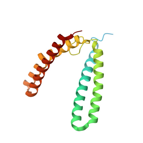
    Rubrerythrins are a group of proteins within the Ferritin-like superfamily that display a defining four-helix bundle domain. They also show multiple structural features that are crucial to their functionality as iron storage proteins and in detoxification and oxidative stress response. Here we investigate rubrerythrin (Rbr) in multiple metalated states, from the pathogen Burkholderia pseudomallei ( Bp ). We use X-ray crystallography for structure determination of Rbr to probe the capacity and specificity of metal binding. Bp Rbr lacks the rubredoxin moiety found in canonical Rbrs from anaerobic lineages, and we demonstrate that Bp Rbr also possesses a domain-swapped dimer, which has functional implications for its putative role in oxidative stress response. We also carry out in crystallo spectroscopic assessment of Bp Rbr with various metals, using energy dispersive X-ray (EDX) spectroscopy. We observe that samples can contain metals other than those supplied in crystallization conditions, and developed a strategy of utilizing EDX spectroscopy to select those samples with single metal incorporation for downstream diffraction data collection. Our work underscores the importance of spectroscopic probing for definitive metal identification and characterization. Present structures of apo-, Fe-, Mn-, and Co-bound rubrerythrin from B. pseudomallei Bp Rbr lacks rubredoxin domain and possesses unique domain swap fold in the dimer Bp Rbr features include promiscuous metal binding and pre-formed metal binding sites Highlights importance of in crystallo spectroscopy in investigating metalloproteins Mutant Rbr with abrogated metal binding supports premise of pre-formed binding sites.

Macromolecule Content 

  • Total Structure Weight: 113.33 kDa 
  • Atom Count: 7,475 
  • Modeled Residue Count: 829 
  • Deposited Residue Count: 1,014 
  • Unique protein chains: 1

Macromolecules

Find similar proteins by:|  3D Structure
Entity ID: 1
MoleculeChains  Sequence LengthOrganismDetailsImage
Rubrerythrin169Burkholderia pseudomalleiMutation(s): 0 
Gene Names: BURPS1710b_A0924
UniProt
Find proteins for Q3JK21 (Burkholderia pseudomallei (strain 1710b))
Explore Q3JK21 
Go to UniProtKB:  Q3JK21
Entity Groups
Sequence Clusters30% Identity50% Identity70% Identity90% Identity95% Identity100% Identity
UniProt GroupQ3JK21
Sequence Annotations
Expand
Reference Sequence

Small Molecules

Ligands 2 Unique
IDChains Name / Formula / InChI Key2D Diagram3D Interactions
PEG

Query on PEG


Download:Ideal Coordinates CCD File
G [auth A]
J [auth B]
K [auth B]
N [auth M]
S [auth Y]
G [auth A],
J [auth B],
K [auth B],
N [auth M],
S [auth Y],
V [auth 5],
W [auth 5]
DI(HYDROXYETHYL)ETHER
C4 H10 O3
MTHSVFCYNBDYFN-UHFFFAOYSA-N
MN

Query on MN


Download:Ideal Coordinates CCD File
H [auth A]
I [auth A]
L [auth B]
M [auth B]
O [auth M]
H [auth A],
I [auth A],
L [auth B],
M [auth B],
O [auth M],
P [auth M],
Q [auth S],
R [auth S],
T [auth Y],
U [auth Y],
X [auth 5],
Y [auth 5]
MANGANESE (II) ION
Mn
WAEMQWOKJMHJLA-UHFFFAOYSA-N

Experimental Data & Validation

Experimental Data

  • Method: X-RAY DIFFRACTION
  • Resolution: 1.58 Å
  • R-Value Free:  0.175 (Depositor), 0.175 (DCC) 
  • R-Value Work:  0.148 (Depositor), 0.148 (DCC) 
Space Group: H 3
Unit Cell:
Length ( Å )Angle ( ˚ )
a = 201.874α = 90
b = 201.874β = 90
c = 69.025γ = 120
Software Package:
Software NamePurpose
REFMACrefinement
XDSdata reduction
STARANISOdata scaling
MOLREPphasing

Structure Validation

View Full Validation Report


Entry History 

& Funding Information

Deposition Data


Funding OrganizationLocationGrant Number
National Science Foundation (NSF, United States)United StatesDBI1231306
Other privateSkarlow Foundation

Revision History  (Full details and data files)

  • Version 1.0: 2024-02-07
    Type: Initial release
  • Version 1.1: 2025-06-25
    Changes: Database references, Structure summary