8FWU | pdb_00008fwu

Structure of the ligand-binding and transmembrane domains of kainate receptor GluK2 in complex with the positive allosteric modulator BPAM344 and competitive antagonist DNQX


Experimental Data Snapshot

  • Method: ELECTRON MICROSCOPY
  • Resolution: 3.18 Å
  • Aggregation State: PARTICLE 
  • Reconstruction Method: SINGLE PARTICLE 

wwPDB Validation 3D Report Full Report

Validation slider image for 8FWU

This is version 1.1 of the entry. See complete history

Literature

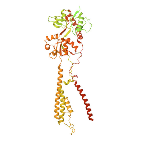
Positive and negative allosteric modulation of GluK2 kainate receptors by BPAM344 and antiepileptic perampanel.

Gangwar, S.P.Yen, L.Y.Yelshanskaya, M.V.Sobolevsky, A.I.

(2023) Cell Rep 42: 112124-112124

  • DOI: https://doi.org/10.1016/j.celrep.2023.112124
  • Primary Citation Related Structures: 
    8FWQ, 8FWR, 8FWS, 8FWT, 8FWU, 8FWV, 8FWW

  • PubMed Abstract: 

    Kainate receptors (KARs) are a subtype of ionotropic glutamate receptors that control synaptic transmission in the central nervous system and are implicated in neurological, psychiatric, and neurodevelopmental disorders. Understanding the regulation of KAR function by small molecules is essential for exploring these receptors as drug targets. Here, we present cryoelectron microscopy (cryo-EM) structures of KAR GluK2 in complex with the positive allosteric modulator BPAM344, competitive antagonist DNQX, and negative allosteric modulator, antiepileptic drug perampanel. Our structures show that two BPAM344 molecules bind per ligand-binding domain dimer interface. In the absence of an agonist or in the presence of DNQX, BPAM344 stabilizes GluK2 in the closed state. The closed state is also stabilized by perampanel, which binds to the ion channel extracellular collar sites located in two out of four GluK2 subunits. The molecular mechanisms of positive and negative allosteric modulation of KAR provide a guide for developing new therapeutic strategies.


  • Organizational Affiliation
    • Department of Biochemistry and Molecular Biophysics, Columbia University, 650 West 168(th) Street, New York, NY 10032, USA.

Macromolecule Content 

  • Total Structure Weight: 428.06 kDa 
  • Atom Count: 14,496 
  • Modeled Residue Count: 1,680 
  • Deposited Residue Count: 3,632 
  • Unique protein chains: 1

Macromolecules

Find similar proteins by:|  3D Structure
Entity ID: 1
MoleculeChains  Sequence LengthOrganismDetailsImage
Glutamate receptor ionotropic, kainate 2
A, B, C, D
908Rattus norvegicusMutation(s): 0 
Gene Names: Grik2Glur6
Membrane Entity: Yes 
UniProt
Find proteins for P42260 (Rattus norvegicus)
Explore P42260 
Go to UniProtKB:  P42260
Entity Groups
Sequence Clusters30% Identity50% Identity70% Identity90% Identity95% Identity100% Identity
UniProt GroupP42260
Glycosylation
Glycosylation Sites: 2Go to GlyGen: P42260-1
Sequence Annotations
Expand
Reference Sequence

Oligosaccharides

Help  
Entity ID: 2
MoleculeChains Length2D Diagram GlycosylationD Interactions
2-acetamido-2-deoxy-beta-D-glucopyranose-(1-4)-2-acetamido-2-deoxy-beta-D-glucopyranose
E, G, H, J
2N-Glycosylation
Glycosylation Resources
GlyTouCan: G42666HT
GlyCosmos: G42666HT
GlyGen: G42666HT
Entity ID: 3
MoleculeChains Length2D Diagram GlycosylationD Interactions
beta-D-mannopyranose-(1-3)-beta-D-mannopyranose-(1-4)-2-acetamido-2-deoxy-beta-D-glucopyranose-(1-4)-2-acetamido-2-deoxy-beta-D-glucopyranose
F, I
4N-Glycosylation
Glycosylation Resources
GlyTouCan: G31886NL
GlyCosmos: G31886NL
GlyGen: G31886NL

Small Molecules

Ligands 6 Unique
IDChains Name / Formula / InChI Key2D Diagram3D Interactions
POV

Query on POV


Download:Ideal Coordinates CCD File
AA [auth B]
FA [auth C]
GA [auth C]
HA [auth C]
IA [auth C]
AA [auth B],
FA [auth C],
GA [auth C],
HA [auth C],
IA [auth C],
KA [auth C],
L [auth A],
LA [auth C],
M [auth A],
MA [auth C],
N [auth A],
O [auth A],
Q [auth A],
R [auth A],
SA [auth D],
Z [auth B]
(2S)-3-(hexadecanoyloxy)-2-[(9Z)-octadec-9-enoyloxy]propyl 2-(trimethylammonio)ethyl phosphate
C42 H82 N O8 P
WTJKGGKOPKCXLL-PFDVCBLKSA-N
DNQ
(Subject of Investigation/LOI)

Query on DNQ


Download:Ideal Coordinates CCD File
EA [auth C],
K [auth A],
RA [auth D],
Y [auth B]
6,7-DINITROQUINOXALINE-2,3-DIONE
C8 H2 N4 O6
YEUPBRRGMWBCEB-UHFFFAOYSA-N
2J9
(Subject of Investigation/LOI)

Query on 2J9


Download:Ideal Coordinates CCD File
BA [auth B],
PA [auth D],
QA [auth D],
X [auth B]
4-cyclopropyl-7-fluoro-3,4-dihydro-2H-1,2,4-benzothiadiazine 1,1-dioxide
C10 H11 F N2 O2 S
FLTMTBPCYAZIKM-UHFFFAOYSA-N
NAG

Query on NAG


Download:Ideal Coordinates CCD File
JA [auth C],
P [auth A]
2-acetamido-2-deoxy-beta-D-glucopyranose
C8 H15 N O6
OVRNDRQMDRJTHS-FMDGEEDCSA-N
CL
(Subject of Investigation/LOI)

Query on CL


Download:Ideal Coordinates CCD File
OA [auth C],
W [auth A]
CHLORIDE ION
Cl
VEXZGXHMUGYJMC-UHFFFAOYSA-M
NA
(Subject of Investigation/LOI)

Query on NA


Download:Ideal Coordinates CCD File
CA [auth B]
DA [auth B]
NA [auth C]
S [auth A]
T [auth A]
CA [auth B],
DA [auth B],
NA [auth C],
S [auth A],
T [auth A],
TA [auth D],
U [auth A],
V [auth A]
SODIUM ION
Na
FKNQFGJONOIPTF-UHFFFAOYSA-N

Experimental Data & Validation

Experimental Data

  • Method: ELECTRON MICROSCOPY
  • Resolution: 3.18 Å
  • Aggregation State: PARTICLE 
  • Reconstruction Method: SINGLE PARTICLE 

Structure Validation

View Full Validation Report


Entry History 

& Funding Information

Deposition Data


Funding OrganizationLocationGrant Number
National Institutes of Health/National Institute of Neurological Disorders and Stroke (NIH/NINDS)United StatesNS083660
National Institutes of Health/National Institute of Neurological Disorders and Stroke (NIH/NINDS)United StatesNS107253

Revision History  (Full details and data files)

  • Version 1.0: 2023-04-19
    Type: Initial release
  • Version 1.1: 2024-10-30
    Changes: Data collection, Structure summary