8FWL | pdb_00008fwl

Crystal structure of Australian bat lyssavirus nucleoprotein in complex with phosphoprotein chaperone


Experimental Data Snapshot

  • Method: X-RAY DIFFRACTION
  • Resolution: 2.19 Å
  • R-Value Free: 
    0.193 (Depositor) 
  • R-Value Work: 
    0.159 (Depositor) 
  • R-Value Observed: 
    0.161 (Depositor) 

Starting Model: experimental
View more details

wwPDB Validation 3D Report Full Report

Validation slider image for 8FWL

This is version 1.2 of the entry. See complete history

Literature

Structural Determination of the Australian Bat Lyssavirus Nucleoprotein and Phosphoprotein Complex.

Donnelly, C.M.Stewart, M.Roby, J.A.Sundaramoorthy, V.Forwood, J.K.

(2023) Viruses 16: 33-33

  • DOI: https://doi.org/10.3390/v16010033
  • Primary Citation Related Structures: 
    8FWL

  • PubMed Abstract: 

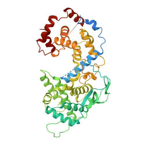
    Australian bat lyssavirus (ABLV) shows similar clinical symptoms as rabies, but there are currently no protein structures available for ABLV proteins. In lyssaviruses, the interaction between nucleoprotein (N) and phosphoprotein (N) in the absence of RNA generates a complex (N 0 P) that is crucial for viral assembly, and understanding the interface between these two proteins has the potential to provide insight into a key feature: the viral lifecycle. In this study, we used recombinant chimeric protein expression and X-ray crystallography to determine the structure of ABLV nucleoprotein bound to residues 1-40 of its phosphoprotein chaperone. Comparison of our results with the recently generated structure of RABV CVS-11 N 0 P demonstrated a highly conserved interface in this complex. Because the N 0 P interface is conserved in the lyssaviruses of phylogroup I, it is an attractive therapeutic target for multiple rabies-causing viral species.


  • Organizational Affiliation
    • School of Dentistry and Medical Sciences, Charles Sturt University, Wagga Wagga, NSW 2678, Australia.

Macromolecule Content 

  • Total Structure Weight: 63.9 kDa 
  • Atom Count: 3,483 
  • Modeled Residue Count: 413 
  • Deposited Residue Count: 567 
  • Unique protein chains: 1

Macromolecules

Find similar proteins by:|  3D Structure
Entity ID: 1
MoleculeChains  Sequence LengthOrganismDetailsImage
Phosphoprotein,Nucleoprotein567Lyssavirus australisMutation(s): 0 
UniProt
Find proteins for Q9YZ24 (Lyssavirus australis)
Explore Q9YZ24 
Go to UniProtKB:  Q9YZ24
Entity Groups
Sequence Clusters30% Identity50% Identity70% Identity90% Identity95% Identity100% Identity
UniProt GroupQ9YZ24
Sequence Annotations
Expand
Reference Sequence

Small Molecules

Ligands 1 Unique
IDChains Name / Formula / InChI Key2D Diagram3D Interactions
PEG

Query on PEG


Download:Ideal Coordinates CCD File
B [auth A]DI(HYDROXYETHYL)ETHER
C4 H10 O3
MTHSVFCYNBDYFN-UHFFFAOYSA-N

Experimental Data & Validation

Experimental Data

  • Method: X-RAY DIFFRACTION
  • Resolution: 2.19 Å
  • R-Value Free:  0.193 (Depositor) 
  • R-Value Work:  0.159 (Depositor) 
  • R-Value Observed: 0.161 (Depositor) 
Space Group: P 1 2 1
Unit Cell:
Length ( Å )Angle ( ˚ )
a = 82.373α = 90
b = 35.503β = 92.63
c = 89.091γ = 90
Software Package:
Software NamePurpose
PHENIXrefinement
XDSdata reduction
Aimlessdata scaling
PHASERphasing
Cootmodel building

Structure Validation

View Full Validation Report


Entry History 

& Funding Information

Deposition Data


Funding OrganizationLocationGrant Number
Not fundedAustralia--

Revision History  (Full details and data files)

  • Version 1.0: 2023-03-08
    Type: Initial release
  • Version 1.1: 2023-03-22
    Changes: Structure summary
  • Version 1.2: 2024-03-20
    Changes: Data collection, Database references