8FPT | pdb_00008fpt

STRUCTURE OF ALPHA-SYNUCLEIN FIBRILS DERIVED FROM HUMAN LEWY BODY DEMENTIA TISSUE


Experimental Data Snapshot

  • Method: SOLID-STATE NMR
  • Conformers Calculated: 256 
  • Conformers Submitted: 10 
  • Selection Criteria: structures with the lowest energy 

wwPDB Validation 3D Report Full Report

Validation slider image for 8FPT

This is version 1.2 of the entry. See complete history

Literature

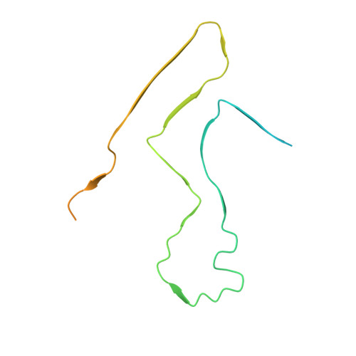
Structure of alpha-synuclein fibrils derived from human Lewy body dementia tissue.

Dhavale, D.D.Barclay, A.M.Borcik, C.G.Basore, K.Berthold, D.A.Gordon, I.R.Liu, J.Milchberg, M.H.O'Shea, J.Y.Rau, M.J.Smith, Z.Sen, S.Summers, B.Smith, J.Warmuth, O.A.Perrin, R.J.Perlmutter, J.S.Chen, Q.Fitzpatrick, J.A.J.Schwieters, C.D.Tajkhorshid, E.Rienstra, C.M.Kotzbauer, P.T.

(2024) Nat Commun 15: 2750-2750

  • DOI: https://doi.org/10.1038/s41467-024-46832-5
  • Primary Citation Related Structures: 
    8FPT

  • PubMed Abstract: 

    The defining feature of Parkinson disease (PD) and Lewy body dementia (LBD) is the accumulation of alpha-synuclein (Asyn) fibrils in Lewy bodies and Lewy neurites. Here we develop and validate a method to amplify Asyn fibrils extracted from LBD postmortem tissue samples and use solid state nuclear magnetic resonance (SSNMR) studies to determine atomic resolution structure. Amplified LBD Asyn fibrils comprise a mixture of single protofilament and two protofilament fibrils with very low twist. The protofilament fold is highly similar to the fold determined by a recent cryo-electron microscopy study for a minority population of twisted single protofilament fibrils extracted from LBD tissue. These results expand the structural characterization of LBD Asyn fibrils and approaches for studying disease mechanisms, imaging agents and therapeutics targeting Asyn.


  • Organizational Affiliation
    • Department of Neurology and Hope Center for Neurological Disorders, Washington University School of Medicine, St. Louis, MO, 63110, USA.

Macromolecule Content 

  • Total Structure Weight: 144.76 kDa 
  • Atom Count: 5,140 
  • Modeled Residue Count: 740 
  • Deposited Residue Count: 1,400 
  • Unique protein chains: 1

Macromolecules

Find similar proteins by:|  3D Structure
Entity ID: 1
MoleculeChains  Sequence LengthOrganismDetailsImage
Alpha-synuclein
A, B, C, D, E
A, B, C, D, E, F, G, H, I, J
140Homo sapiensMutation(s): 0 
Gene Names: SNCANACPPARK1
UniProt & NIH Common Fund Data Resources
Find proteins for P37840 (Homo sapiens)
Explore P37840 
Go to UniProtKB:  P37840
PHAROS:  P37840
GTEx:  ENSG00000145335 
Entity Groups
Sequence Clusters30% Identity50% Identity70% Identity90% Identity95% Identity100% Identity
UniProt GroupP37840
Sequence Annotations
Expand
Reference Sequence

Experimental Data & Validation

Experimental Data

  • Method: SOLID-STATE NMR
  • Conformers Calculated: 256 
  • Conformers Submitted: 10 
  • Selection Criteria: structures with the lowest energy 

Structure Validation

View Full Validation Report


Entry History 

& Funding Information

Deposition Data


Funding OrganizationLocationGrant Number
National Institutes of Health/National Institute of General Medical Sciences (NIH/NIGMS)United States--

Revision History  (Full details and data files)

  • Version 1.0: 2023-02-22
    Type: Initial release
  • Version 1.1: 2024-05-15
    Changes: Data collection, Database references
  • Version 1.2: 2024-12-11
    Changes: Data collection, Database references, Structure summary