8FO6

Nucleotide-free structure of a functional construct of eukaryotic elongation factor 2 kinase.


Experimental Data Snapshot

  • Method: X-RAY DIFFRACTION
  • Resolution: 2.55 Å
  • R-Value Free: 0.231 
  • R-Value Work: 0.217 
  • R-Value Observed: 0.218 

wwPDB Validation   3D Report Full Report


This is version 1.0 of the entry. See complete history


Literature

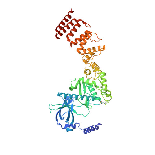
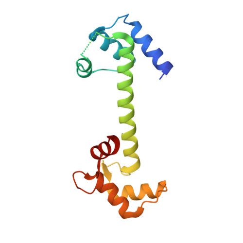
ADP enhances the allosteric activation of eukaryotic elongation factor 2 kinase by calmodulin.

Piserchio, A.Long, K.J.Browning, L.S.Bohanon, A.L.Isiorho, E.A.Dalby, K.N.Ghose, R.

(2023) Proc Natl Acad Sci U S A 120: e2300902120-e2300902120

  • DOI: https://doi.org/10.1073/pnas.2300902120
  • Primary Citation of Related Structures:  
    8FNY, 8FO6

  • PubMed Abstract: 

    Protein translation, one of the most energy-consumptive processes in a eukaryotic cell, requires robust regulation, especially under energy-deprived conditions. A critical component of this regulation is the suppression of translational elongation through reduced ribosome association of the GTPase eukaryotic elongation factor 2 (eEF-2) resulting from its specific phosphorylation by the calmodulin (CaM)-activated α-kinase eEF-2 kinase (eEF-2K). It has been suggested that the eEF-2K response to reduced cellular energy levels is indirect and mediated by the universal energy sensor AMP-activated protein kinase (AMPK) through direct stimulatory phosphorylation and/or downregulation of the eEF-2K-inhibitory nutrient-sensing mTOR pathway. Here, we provide structural, biochemical, and cell-biological evidence of a direct energy-sensing role of eEF-2K through its stimulation by ADP. A crystal structure of the nucleotide-bound complex between CaM and the functional core of eEF-2K phosphorylated at its primary stimulatory site (T348) reveals ADP bound at a unique pocket located on the face opposite that housing the kinase active site. Within this basic pocket (BP), created at the CaM/eEF-2K interface upon complex formation, ADP is stabilized through numerous interactions with both interacting partners. Biochemical analyses using wild-type eEF-2K and specific BP mutants indicate that ADP stabilizes CaM within the active complex, increasing the sensitivity of the kinase to CaM. Induction of energy stress through glycolysis inhibition results in significantly reduced enhancement of phosphorylated eEF-2 levels in cells expressing ADP-binding compromised BP mutants compared to cells expressing wild-type eEF-2K. These results suggest a direct energy-sensing role for eEF-2K through its cooperative interaction with CaM and ADP.


  • Organizational Affiliation

    Department of Chemistry and Biochemistry, The City College of New York, New York, NY 10031.


Macromolecules
Find similar proteins by:  (by identity cutoff)  |  3D Structure
Entity ID: 1
MoleculeChains Sequence LengthOrganismDetailsImage
Eukaryotic elongation factor 2 kinase531Homo sapiensMutation(s): 0 
Gene Names: EEF2K
EC: 2.7.11.20
UniProt & NIH Common Fund Data Resources
Find proteins for O00418 (Homo sapiens)
Explore O00418 
Go to UniProtKB:  O00418
PHAROS:  O00418
GTEx:  ENSG00000103319 
Entity Groups  
Sequence Clusters30% Identity50% Identity70% Identity90% Identity95% Identity100% Identity
UniProt GroupO00418
Sequence Annotations
Expand
  • Reference Sequence
Find similar proteins by:  (by identity cutoff)  |  3D Structure
Entity ID: 2
MoleculeChains Sequence LengthOrganismDetailsImage
Calmodulin-1148Homo sapiensMutation(s): 0 
Gene Names: CALM1CALMCAMCAM1
UniProt & NIH Common Fund Data Resources
Find proteins for P0DP23 (Homo sapiens)
Explore P0DP23 
Go to UniProtKB:  P0DP23
PHAROS:  P0DP23
GTEx:  ENSG00000198668 
Entity Groups  
Sequence Clusters30% Identity50% Identity70% Identity90% Identity95% Identity100% Identity
UniProt GroupP0DP23
Sequence Annotations
Expand
  • Reference Sequence
Small Molecules
Modified Residues  1 Unique
IDChains TypeFormula2D DiagramParent
TPO
Query on TPO
A
L-PEPTIDE LINKINGC4 H10 N O6 PTHR
Experimental Data & Validation

Experimental Data

  • Method: X-RAY DIFFRACTION
  • Resolution: 2.55 Å
  • R-Value Free: 0.231 
  • R-Value Work: 0.217 
  • R-Value Observed: 0.218 
  • Space Group: P 1 21 1
Unit Cell:
Length ( Å )Angle ( ˚ )
a = 77.527α = 90
b = 58.901β = 109.67
c = 88.394γ = 90
Software Package:
Software NamePurpose
autoPROCdata processing
Aimlessdata scaling
TRUNCATEdata processing
PHENIXrefinement
XDSdata reduction
PHASERphasing
ISOLDErefinement
Cootmodel building

Structure Validation

View Full Validation Report


Entry History & Funding Information

Deposition Data


Funding OrganizationLocationGrant Number
National Institutes of Health/National Institute of General Medical Sciences (NIH/NIGMS)United StatesGM123252

Revision History  (Full details and data files)

  • Version 1.0: 2023-05-03
    Type: Initial release