


Crystal Structures of Diaryl Hydrazone and Sulfone Stabilizers in Complex with an Amyloidogenic Light Chain Reveal an Alternate Ligand-Binding Cavity
Yan, N.L., Wilson, I.A., Kelly, J.W.(2023) Isr J Chem
Experimental Data Snapshot
Starting Model: experimental
View more details
(2023) Isr J Chem
Macromolecule Content
Entity ID: 1 | |||||
|---|---|---|---|---|---|
| Molecule | Chains | Sequence Length | Organism | Details | Image |
| H9 immunoglobulin light chain | 216 | Homo sapiens | Mutation(s): 0 | 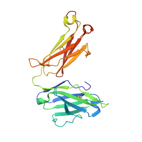 | |
Entity Groups | |||||
| Sequence Clusters | 30% Identity50% Identity70% Identity90% Identity95% Identity100% Identity | ||||
Sequence AnnotationsExpand | |||||
Reference Sequence | |||||
| Ligands 2 Unique | |||||
|---|---|---|---|---|---|
| ID | Chains | Name / Formula / InChI Key | 2D Diagram | 3D Interactions | |
| Y4K (Subject of Investigation/LOI) Download:Ideal Coordinates CCD File | M [auth A] P [auth C] Q [auth G] T [auth I] U [auth E] | 3-nitro-4-{2-[(2S)-2-phenylpropyl]hydrazin-1-yl}benzene-1-sulfonamide C15 H18 N4 O4 S NSSXWTMELVHXJK-LLVKDONJSA-N |  | ||
| PO4 Download:Ideal Coordinates CCD File | N [auth A] O [auth B] R [auth G] S [auth H] V [auth E] | PHOSPHATE ION O4 P NBIIXXVUZAFLBC-UHFFFAOYSA-K |  | ||
| Length ( Å ) | Angle ( ˚ ) |
|---|---|
| a = 63.141 | α = 106.31 |
| b = 95.655 | β = 92.93 |
| c = 125.918 | γ = 90.16 |
| Software Name | Purpose |
|---|---|
| REFMAC | refinement |
| XDS | data reduction |
| SCALA | data scaling |
| PHASER | phasing |
| Funding Organization | Location | Grant Number |
|---|---|---|
| National Institutes of Health/National Heart, Lung, and Blood Institute (NIH/NHLBI) | United States | HL157566 |
| National Institutes of Health/National Heart, Lung, and Blood Institute (NIH/NHLBI) | United States | HL154732 |


