8FMH

Structure of CBASS Cap5 from Pseudomonas syringae as an activated tetramer with the cyclic dinucleotide 3'2'-c-dGAMP ligand (2 tetramers in the AU)


Experimental Data Snapshot

  • Method: X-RAY DIFFRACTION
  • Resolution: 1.87 Å
  • R-Value Free: 0.253 
  • R-Value Work: 0.210 
  • R-Value Observed: 0.212 

wwPDB Validation   3D Report Full Report


Ligand Structure Quality Assessment 


This is version 1.1 of the entry. See complete history


Literature

Activation of CBASS Cap5 endonuclease immune effector by cyclic nucleotides.

Rechkoblit, O.Sciaky, D.Kreitler, D.F.Buku, A.Kottur, J.Aggarwal, A.K.

(2024) Nat Struct Mol Biol 

  • DOI: https://doi.org/10.1038/s41594-024-01220-x
  • Primary Citation of Related Structures:  
    8FM1, 8FMF, 8FMG, 8FMH

  • PubMed Abstract: 

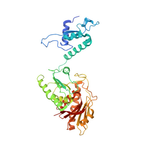
    The bacterial cyclic oligonucleotide-based antiphage signaling system (CBASS) is similar to the cGAS-STING system in humans, containing an enzyme that synthesizes a cyclic nucleotide on viral infection and an effector that senses the second messenger for the antiviral response. Cap5, containing a SAVED domain coupled to an HNH DNA endonuclease domain, is the most abundant CBASS effector, yet the mechanism by which it becomes activated for cell killing remains unknown. We present here high-resolution structures of full-length Cap5 from Pseudomonas syringae (Ps) with second messengers. The key to PsCap5 activation is a dimer-to-tetramer transition, whereby the binding of second messenger to dimer triggers an open-to-closed transformation of the SAVED domains, furnishing a surface for assembly of the tetramer. This movement propagates to the HNH domains, juxtaposing and converting two HNH domains into states for DNA destruction. These results show how Cap5 effects bacterial cell suicide and we provide proof-in-principle data that the CBASS can be extrinsically activated to limit bacterial infections.


  • Organizational Affiliation

    Department of Pharmacological Sciences, Icahn School of Medicine at Mount Sinai, New York, NY, USA. olga.rechkoblit@mssm.edu.


Macromolecules
Find similar proteins by:  (by identity cutoff)  |  3D Structure
Entity ID: 1
MoleculeChains Sequence LengthOrganismDetailsImage
SAVED domain-containing protein
A, B, C, D, E
A, B, C, D, E, F, G, H
388Pseudomonas syringaeMutation(s): 0 
UniProt
Find proteins for A0A2P0QGK5 (Pseudomonas syringae pv. actinidiae)
Explore A0A2P0QGK5 
Go to UniProtKB:  A0A2P0QGK5
Entity Groups  
Sequence Clusters30% Identity50% Identity70% Identity90% Identity95% Identity100% Identity
UniProt GroupA0A2P0QGK5
Sequence Annotations
Expand
  • Reference Sequence
Small Molecules
Ligands 6 Unique
IDChains Name / Formula / InChI Key2D Diagram3D Interactions
4UR (Subject of Investigation/LOI)
Query on 4UR

Download Ideal Coordinates CCD File 
AA [auth F]
DA [auth G]
I [auth A]
JA [auth H]
M [auth B]
AA [auth F],
DA [auth G],
I [auth A],
JA [auth H],
M [auth B],
P [auth C],
U [auth D],
W [auth E]
3'2'-cGAMP
C20 H24 N10 O13 P2
FAFONCPHZLORMH-INFSMZHSSA-N
PGE
Query on PGE

Download Ideal Coordinates CCD File 
Q [auth C]TRIETHYLENE GLYCOL
C6 H14 O4
ZIBGPFATKBEMQZ-UHFFFAOYSA-N
PEG
Query on PEG

Download Ideal Coordinates CCD File 
EA [auth G]DI(HYDROXYETHYL)ETHER
C4 H10 O3
MTHSVFCYNBDYFN-UHFFFAOYSA-N
GOL
Query on GOL

Download Ideal Coordinates CCD File 
FA [auth G]GLYCEROL
C3 H8 O3
PEDCQBHIVMGVHV-UHFFFAOYSA-N
ZN
Query on ZN

Download Ideal Coordinates CCD File 
BA [auth F]
GA [auth G]
J [auth A]
KA [auth H]
N [auth B]
BA [auth F],
GA [auth G],
J [auth A],
KA [auth H],
N [auth B],
R [auth C],
V [auth D],
X [auth E]
ZINC ION
Zn
PTFCDOFLOPIGGS-UHFFFAOYSA-N
MG
Query on MG

Download Ideal Coordinates CCD File 
CA [auth F]
HA [auth G]
IA [auth G]
K [auth A]
L [auth A]
CA [auth F],
HA [auth G],
IA [auth G],
K [auth A],
L [auth A],
O [auth B],
S [auth C],
T [auth C],
Y [auth E],
Z [auth E]
MAGNESIUM ION
Mg
JLVVSXFLKOJNIY-UHFFFAOYSA-N
Experimental Data & Validation

Experimental Data

  • Method: X-RAY DIFFRACTION
  • Resolution: 1.87 Å
  • R-Value Free: 0.253 
  • R-Value Work: 0.210 
  • R-Value Observed: 0.212 
  • Space Group: P 1 21 1
Unit Cell:
Length ( Å )Angle ( ˚ )
a = 67.97α = 90
b = 295.678β = 113.79
c = 84.052γ = 90
Software Package:
Software NamePurpose
PHENIXrefinement
PHENIXrefinement
XDSdata reduction
Aimlessdata scaling
PHASERphasing

Structure Validation

View Full Validation Report


Ligand Structure Quality Assessment 


Entry History & Funding Information

Deposition Data


Funding OrganizationLocationGrant Number
National Institutes of Health/National Institute of General Medical Sciences (NIH/NIGMS)United StatesGM13170

Revision History  (Full details and data files)

  • Version 1.0: 2024-02-07
    Type: Initial release
  • Version 1.1: 2024-02-21
    Changes: Database references