8FJE

The five-repeat design E8


Experimental Data Snapshot

  • Method: X-RAY DIFFRACTION
  • Resolution: 3.00 Å
  • R-Value Free: 0.299 
  • R-Value Work: 0.267 
  • R-Value Observed: 0.271 

wwPDB Validation   3D Report Full Report


This is version 1.3 of the entry. See complete history


Literature

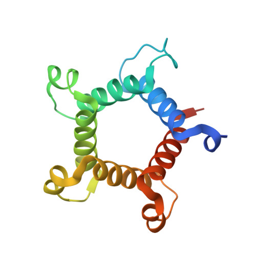
Hallucination of closed repeat proteins containing central pockets.

An, L.Hicks, D.R.Zorine, D.Dauparas, J.Wicky, B.I.M.Milles, L.F.Courbet, A.Bera, A.K.Nguyen, H.Kang, A.Carter, L.Baker, D.

(2023) Nat Struct Mol Biol 30: 1755-1760

  • DOI: https://doi.org/10.1038/s41594-023-01112-6
  • Primary Citation of Related Structures:  
    8FJE, 8FJF, 8FJG

  • PubMed Abstract: 

    In pseudocyclic proteins, such as TIM barrels, β barrels, and some helical transmembrane channels, a single subunit is repeated in a cyclic pattern, giving rise to a central cavity that can serve as a pocket for ligand binding or enzymatic activity. Inspired by these proteins, we devised a deep-learning-based approach to broadly exploring the space of closed repeat proteins starting from only a specification of the repeat number and length. Biophysical data for 38 structurally diverse pseudocyclic designs produced in Escherichia coli are consistent with the design models, and the three crystal structures we were able to obtain are very close to the designed structures. Docking studies suggest the diversity of folds and central pockets provide effective starting points for designing small-molecule binders and enzymes.


  • Organizational Affiliation

    Department of Biochemistry, University of Washington, Seattle, WA, USA. linnaan@uw.edu.


Macromolecules
Find similar proteins by:  (by identity cutoff)  |  3D Structure
Entity ID: 1
MoleculeChains Sequence LengthOrganismDetailsImage
E8
A, B
145synthetic constructMutation(s): 0 
Entity Groups  
Sequence Clusters30% Identity50% Identity70% Identity90% Identity95% Identity100% Identity
Sequence Annotations
Expand
  • Reference Sequence
Experimental Data & Validation

Experimental Data

  • Method: X-RAY DIFFRACTION
  • Resolution: 3.00 Å
  • R-Value Free: 0.299 
  • R-Value Work: 0.267 
  • R-Value Observed: 0.271 
  • Space Group: P 2 21 21
Unit Cell:
Length ( Å )Angle ( ˚ )
a = 47.859α = 90
b = 65.763β = 90
c = 87.434γ = 90
Software Package:
Software NamePurpose
PHENIXrefinement
XDSdata reduction
XSCALEdata scaling
PHASERphasing

Structure Validation

View Full Validation Report


Entry History & Funding Information

Deposition Data


Funding OrganizationLocationGrant Number
Howard Hughes Medical Institute (HHMI)United States--

Revision History  (Full details and data files)

  • Version 1.0: 2023-09-27
    Type: Initial release
  • Version 1.1: 2023-10-04
    Changes: Database references, Structure summary
  • Version 1.2: 2023-10-11
    Changes: Database references, Refinement description
  • Version 1.3: 2023-11-22
    Changes: Database references