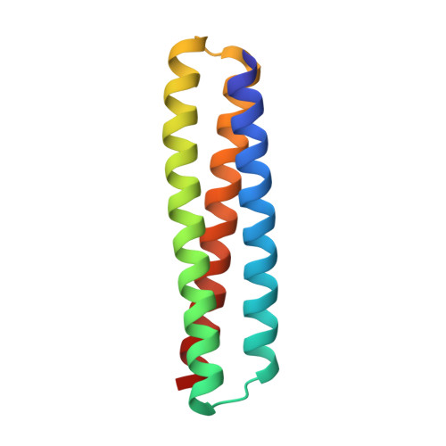
Design of stimulus-responsive two-state hinge proteins.
Praetorius, F., Leung, P.J.Y., Tessmer, M.H., Broerman, A., Demakis, C., Dishman, A.F., Pillai, A., Idris, A., Juergens, D., Dauparas, J., Li, X., Levine, P.M., Lamb, M., Ballard, R.K., Gerben, S.R., Nguyen, H., Kang, A., Sankaran, B., Bera, A.K., Volkman, B.F., Nivala, J., Stoll, S., Baker, D.(2023) Science 381: 754-760
- PubMed: 37590357 Search on PubMedSearch on PubMed Central
- DOI: https://doi.org/10.1126/science.adg7731
- Primary Citation Related Structures:
8FIH, 8FIN, 8FIQ, 8FIT, 8FVT - PubMed Abstract:
In nature, proteins that switch between two conformations in response to environmental stimuli structurally transduce biochemical information in a manner analogous to how transistors control information flow in computing devices. Designing proteins with two distinct but fully structured conformations is a challenge for protein design as it requires sculpting an energy landscape with two distinct minima. Here we describe the design of "hinge" proteins that populate one designed state in the absence of ligand and a second designed state in the presence of ligand. X-ray crystallography, electron microscopy, double electron-electron resonance spectroscopy, and binding measurements demonstrate that despite the significant structural differences the two states are designed with atomic level accuracy and that the conformational and binding equilibria are closely coupled.
- Department of Biochemistry, University of Washington, Seattle, WA, USA.
Organizational Affiliation: